
alleen voor onderzoeksdoeleinden
Y-27632 Dihydrochloride ROCK remmer
Cat.nr.S1049
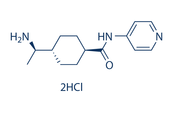
Chemische structuur
Molecuulgewicht: 320.26
Kwaliteitscontrole
Producten die vaak samen worden gebruikt met Y-27632 Dihydrochloride
| Gerelateerde doelwitten | CDK HSP PD-1/PD-L1 Wee1 DNA/RNA Synthesis Microtubule Associated Ras KRas Aurora Kinase Casein Kinase |
|---|---|
| Overig ROCK Inhibitoren | Thiazovivin (TZV) Y-27632 NRL-1049 Ripasudil hydrochloride dihydrate Fasudil (HA-1077) Hydrochloride GSK429286A RKI-1447 GSK269962A HCl Y-39983 Dihydrochloride Hydroxyfasudil HCl |
Celkweek, behandeling & werkzame concentratie
| Cellijnen | Assaytype | Concentratie | Incubatietijd | Formulering | Activiteitsbeschrijving | PMID |
|---|---|---|---|---|---|---|
| Salivary gland stem cells | Function Assay | 10 µM | 7 d | Reduces SGSC senescence | 25804560 | |
| HT22 | Cytotoxic Assay | 10 µM | 13 h | Protects against glutamate-induced neuronal death | 22810835 | |
| Swiss3T3 | Colony-forming Assay | 10 µM | 13 d | Increases prostate cell colony-forming activity | 21464902 | |
| TSGH 8301 | Migration Assay | 20 µM | 1 h | Increases cell migration | 19896475 | |
| Cynomolgus monkey embryonic stem cells | Cytotoxic Assay | 20 µM | 24 h | Promotes cyES cell survival | 18940855 | |
| PC 12 | Function Assay | 10 μM | 24 h | Attenuates catecholamine biosynthesis | 16219424 | |
| C2C12 | Function Assay | 10 μM | 6 h | Prevents the serine phosphorylation of IRS-1 induced by insulin and/or TNF-α | 16267124 | |
| Pancreatic acinar cells | Function Assay | 10 μM | 70 min | Potentiates CCK-stimulated pancreatic enzyme secretion | 12745080 | |
| Rat hepatic stellate cells | Function Assay | 30 μM | 48 h | Diminishes the phosphorylation of Erk2, and decreases new DNA synthesis | 10845663 | |
| LNCaP | Growth Inhibition Assay | 25 μM | 17 h | Does not inhibit cell growth | 10720471 | |
| PC3 | Growth Inhibition Assay | 25 μM | 17 h | Does not inhibit cell growth | 10720471 | |
| PC3 | Migration Assay | 25 μM | 1 h | Inhibits the BMFB-CM and the EGF-stimulated migration | 10720471 | |
| PC3 | Function Assay | 25 μM | 1 h | Induces morphological changes | 10720471 | |
| Rat Vascular Smooth Muscle Cells | Function Assay | 10 μM | 2 h | Inhibits angiotensin II-induced hypertrophy | 10642317 | |
| Stellate Cell | Function Assay | 25 μM | 15 min | Inhibits formation of F-actin stress fibers and phosphorylation of myosin light chain | 10600496 | |
| Neonatal rat ventricular myocytes | Function Assay | 10 μM | 48 h | Inhibits ET-1-induced increases in protein synthesis, cell size and myofibrillar organization | 10386613 | |
| LS174T | Growth Inhibition Assay | 10 μM | 18 d | Moderately inhibits cell growth | 10021386 | |
| HCT116 | Growth Inhibition Assay | 10 μM | 18 d | Strongly inhibits cell growth | 10021386 | |
| HCT15 | Growth Inhibition Assay | 10 μM | 18 d | Does not inhibit cell growth | 10021386 | |
| SW620 | Growth Inhibition Assay | 10 μM | 18 d | Does not inhibit cell growth | 10021386 | |
| Src-2 | Growth Inhibition Assay | 10 μM | 18 d | Does not inhibit cell growth | 10021386 | |
| Src-1 | Growth Inhibition Assay | 10 μM | 18 d | Does not inhibit cell growth | 10021386 | |
| NIH3T3 | Growth Inhibition Assay | 10 μM | 18 d | Does not inhibit cell growth | 10021386 | |
| Src-4 | Growth Inhibition Assay | 10 μM | 18 d | Does not inhibit cell growth | 10021386 | |
| Src-1 | Growth Inhibition Assay | 10 μM | 18 d | Does not inhibit cell growth | 10021386 | |
| Ras-4 | Growth Inhibition Assay | 10 μM | 18 d | Strongly inhibits cell growth | 10021386 | |
| Ras-2 | Growth Inhibition Assay | 10 μM | 18 d | Strongly inhibits cell growth | 10021386 | |
| mNET1-e | Growth Inhibition Assay | 10 μM | 18 d | Strongly inhibits cell growth | 10021386 | |
| mNET1-d | Growth Inhibition Assay | 10 μM | 18 d | Strongly inhibits cell growth | 10021386 | |
| Dbl-e | Growth Inhibition Assay | 10 μM | 18 d | Moderately inhibits cell growth | 10021386 | |
| Dbl-d | Growth Inhibition Assay | 10 μM | 18 d | Strongly inhibits cell growth | 10021386 | |
| NIH3T3 | Growth Inhibition Assay | 10 μM | 18 d | Does not inhibit cell growth | 10021386 | |
| Mesothelial cells from rat mesentery | Invasive Assay | 30 μM | 20 h | Blocks invasive activity | 9930872 | |
| CCL39 | Function Assay | 30 μM | 30 min | Completely abolishes activation of Na-H exchanger NHE1 by integrins | 9693382 | |
| HeLa | Function Assay | 10 μM | 30 min | Inhibits the formation of stress fibers and the assembly of vinculin-containing focal adhesions | 9668072 | |
| N1E-115 | Function Assay | 10 μM | 2 h | DMSO | Inhibits the assembly of microtubules and intermediate filaments to form extended processes | 9647654 |
| Swiss 3T3 cells | Function Assay | 10 μM | 2 h | DMSO | Inhibits the assembly of microtubules and intermediate filaments to form extended processes | 9647654 |
| Klik om meer experimentele gegevens over cellijnen te bekijken | ||||||
Chemische informatie, opslag en stabiliteit
| Molecuulgewicht | 320.26 | Formule | C14H21N3O.2HCl |
Opslag (vanaf de datum van ontvangst) | |
|---|---|---|---|---|---|
| CAS-nr. | 129830-38-2 | SDF downloaden | Opslag van stamoplossingen |
|
|
| Synoniemen | N/A | Smiles | CC(C1CCC(CC1)C(=O)NC2=CC=NC=C2)N.Cl.Cl | ||
Oplosbaarheid
|
In vitro |
DMSO
: 64 mg/mL
(199.83 mM)
Water : 64 mg/mL Ethanol : 4 mg/mL |
Molariteitscalculator
|
In vivo |
|||||
In vivo formulatiecalculator (heldere oplossing)
Stap 1: Voer onderstaande informatie in (Aanbevolen: een extra dier om rekening te houden met verlies tijdens het experiment)
Stap 2: Voer de in vivo formulering in (Dit is alleen de calculator, geen formulering. Neem eerst contact met ons op als er geen in vivo formulering is in de sectie oplosbaarheid.)
Berekeningsresultaten:
Werkconcentratie: mg/ml;
Methode voor het bereiden van DMSO-moedervloeistof: mg geneesmiddel vooropgelost in μL DMSO ( Concentratie moedervloeistof mg/mL, Neem eerst contact met ons op als de concentratie de DMSO-oplosbaarheid van de batch van het geneesmiddel overschrijdt. )
Methode voor het bereiden van in vivo formulering: Neem μL DMSO moedervloeistof, voeg daarna toeμL PEG300, mengen en verhelderen, daarna toevoegenμL Tween 80, mengen en verhelderen, daarna toevoegen μL ddH2O, mengen en verhelderen.
Methode voor het bereiden van in vivo formulering: Neem μL DMSO moedervloeistof, voeg daarna toe μL Maïsolie, mengen en verhelderen.
Opmerking: 1. Zorg ervoor dat de vloeistof helder is voordat u het volgende oplosmiddel toevoegt.
2. Zorg ervoor dat u het/de oplosmiddel(en) in de juiste volgorde toevoegt. U moet ervoor zorgen dat de verkregen oplossing, bij de vorige toevoeging, een heldere oplossing is voordat u verdergaat met het toevoegen van het volgende oplosmiddel. Fysieke methoden zoals vortexen, ultrasoon of een warmwaterbad kunnen worden gebruikt om het oplossen te bevorderen.
Werkingsmechanisme
| Targets/IC50/Ki |
ROCK1 (p160ROCK)
(Cell-free assay) 140 nM(Ki)
ROCK2
(Cell-free assay) 300 nM(Ki)
|
|---|---|
| In vitro |
Y-27632 2HCl remt ROCK-II en vertoont weinig activiteit tegen PKC, cAMP-afhankelijke proteïnekinase en myosine lichte-keten kinase (MLCK) met een Ki van respectievelijk 26 μM, 25 μM en > 250 μM, evenals PKA geactiveerd door een ander Rho-familie GTPase-lid, Cdc42. Deze verbinding remt de samentrekking van gladde spieren, geïnduceerd door verschillende agonisten, waaronder fenylefrine, histamine, acetylcholine, serotonine, endotheline en tromboxaan, met een IC50 van 0,3-1 μM, door selectieve remming van Ca2+-sensibilisatie. Het onderdrukt Rho-geïnduceerde, p160ROCK-gemedieerde vorming van stressvezels in gekweekte cellen. Deze chemische behandeling blokkeert zowel Rho-gemedieerde activering van actomyosine als LPA-gestimuleerde invasieve activiteit van MM1-cellen op een concentratieafhankelijke manier. Deze behandeling met deze verbinding is niet alleen voldoende om de vorming van uitbundige axonale processen te initiëren, maar vergemakkelijkt ook de axonale rijping tijdens de allereerste stadia van axonogenese, terwijl de axonale elongatie grotendeels wordt gespaard. In menselijke embryonale stamcellen (hES-cellen) vermindert deze chemische behandeling bij 10 μM de dissociatie-geïnduceerde apoptose aanzienlijk, zelfs in serumvrije suspensiekweek (SFEB), verhoogt de kloneringsefficiëntie (van ~1% tot ~27%), vergemakkelijkt subklonering na genoverdracht, en stelt SFEB-gekweekte hES-cellen in staat om te overleven en te differentiëren tot Bf1+ corticale en basale telencefale voorlopercellen. |
| Kinase Assay |
Fosforyleringsreacties
|
|
De p160ROCK wordt in COS-cellen tot getagde full-length eiwitten gebracht en ge
duceerd met behulp van anti-tag antilichamen. De p160ROCK (30 ng) wordt bij 30 °C in een totaal volume van 31 μL ge
ïncubeerd met 40 μM [γ-32P]ATP (3,3 Ci/mmol) en met 3 μg van ofwel histon (HF2A), gedephosphoryleerd case
\n
ïne of MBP in de aanwezigheid van verschillende concentraties van deze verbinding. Een alicuot van 7 μL wordt genomen bij 0, 5, 10 en 20 minuten, gemengd met een gelijk volume van 2 × Laemmli monsterbuffer en toegepast op SDS-PAGE. De gel wordt gekleurd met Commassie Blue, gedroogd en onderworpen aan analyse door een Bioimage Analyzer BAS2000. De concentratie van deze chemische stof die nodig is om de p160ROCK-activiteit met 50% te remmen (IC50-waarde) wordt verkregen. De Ki-waarde wordt berekend volgens de vergelijking, Ki = IC50/(1 + S/Km), waarbij S en Km de concentraties en Km-waarde voor ATP vertegenwoordigen.
|
|
| In vivo |
Orale toediening van Y-27632 Dihydrochloride (30 mg/kg) verlaagt de bloeddruk significant op een dosisafhankelijke manier bij spontaan hypertensieve ratten, renaal hypertensieve ratten en deoxycorticosteronacetaat (DOCA)-zout hypertensieve ratten. Wanneer deze verbinding gedurende 11 dagen continu wordt toegediend met een snelheid van 0,55 μL per uur via geïmplanteerde pompen, wordt de invasie van tumorcellen (MM1-cellen die Val14-RhoA tot expressie brengen bij ratten) significant vertraagd. Door ROCK te remmen, vermindert deze chemische behandeling hypoxie-geïnduceerde angiogenese en vasculaire remodellering in de pulmonaire circulatie. Voorbehandeling met deze verbinding heeft een beschermend effect tegen tumorvorming bij albinomuizen met Ehrlich-ascitescarcinoom. |
Referenties |
|
Toepassingen
| Methoden | Biomarkers | Afbeeldingen | PMID |
|---|---|---|---|
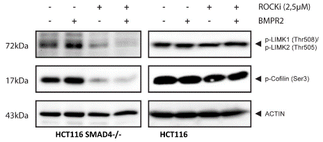
| Western Blot | p-LIMK1(Thr508) p-LIMK2(Thr505) p-Cofilin(Ser3) CDK2 KRT7 ROCK2 E-cadherin p-MLC2(Ser19) |
|
24704720 |
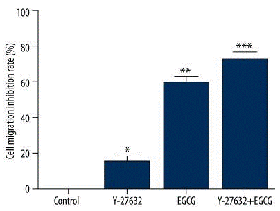
| Transwell migration assay | cell migration inhibition |
|
27694793 |
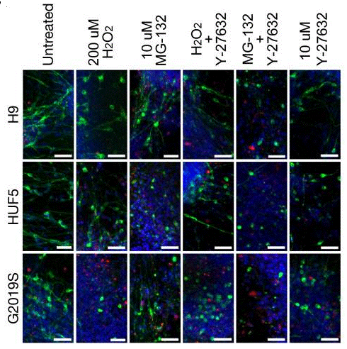
| Immunofluorescence | Neurotoxicity Assay E-cadherin beta-catenin Phalloidin |
|
21362567 |
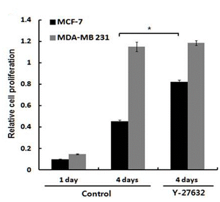
| Growth inhibition assay | cell proliferation |
|
24523903 |
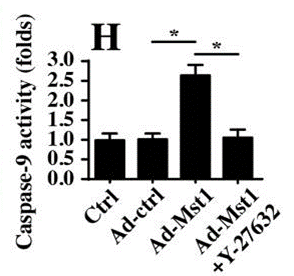
| ELISA | caspase-9 |
|
30320378 |
Technische ondersteuning
Tel: +1-832-582-8158 Ext:3
Als u nog andere vragen heeft, laat dan een bericht achter.
Veelgestelde vragen
Vraag 1:
Is there any data about the Amax (maximum attraction luminosity) and extinction coefficient of it?
Antwoord:
The wavelength used to test its HPLC is 260nm, while the extinction coefficient is unknown.
Vraag 2:
Could it be used in cell culture? Do you have any reference for this application?
Antwoord:
Yes. It can be used in cell culture certainly. Here is the reference website: http://molpharm.aspetjournals.org/content/57/5/976.full.

Producten zijn uitsluitend voor onderzoeksdoeleinden. Niet voor menselijk gebruik. Wij verkopen niet aan patiënten.
©Copyright 2013 Selleck Chemicals. Alle rechten voorbehouden.