alleen voor onderzoeksdoeleinden
Alvespimycin (17-DMAG) Hydrochloride HSP remmer
Cat.nr.S1142
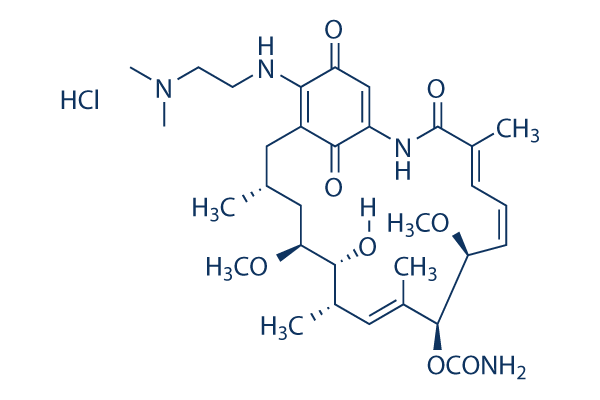
Chemische structuur
Molecuulgewicht: 653.21
Kwaliteitscontrole
| Gerelateerde doelwitten | Akt Wnt/beta-catenin PKC ROCK Microtubule Associated Integrin Bcr-Abl Actin FAK Kinesin |
|---|---|
| Overig HSP Inhibitoren | Tanespimycin (17-AAG) Elesclomol (STA-4783) Luminespib (NVP-AUY922) Ganetespib (STA-9090) VER-155008 Onalespib (AT13387) BIIB021 Zelavespib (PU-H71) HSP990 (NVP-HSP990) NVP-BEP800 |
Celkweek, behandeling & werkzame concentratie
| Cellijnen | Assaytype | Concentratie | Incubatietijd | Formulering | Activiteitsbeschrijving | PMID |
|---|---|---|---|---|---|---|
| MDA-MB-231 | Function assay | Inhibition of Hsp90 in human MDA-MB-231 cells assessed as her2 degradation, IC50=0.0045μM. | 18929486 | |||
| A2058 | Cytotoxicity assay | Cytotoxicity against human A2058 cells by MTT assay, IC50=0.0021μM. | 18929486 | |||
| AGS | Cytotoxicity assay | Cytotoxicity against human AGS cells by MTT assay, IC50=16μM. | 18359631 | |||
| HeLa | Cytotoxicity assay | Cytotoxicity against human HeLa cells by MTT assay, IC50=2.06μM. | 18359631 | |||
| HeLa | Function assay | Inhibition of TNF-alpha-induced NF-kappaB activation in human HeLa cells, IC50=0.15μM. | 18359631 | |||
| AGS | Function assay | Inhibition of hypoxia-induced HIF1 activation in human AGS cells by reporter gene assay, IC50=0.0036μM. | 18359631 | |||
| NCI-H526 | Function assay | 1 uM | 96 hrs | Inhibition of HSP90-mediated proliferation of human NCI-H526 cells at 1 uM after 96 hrs by sulforhodamine B assay | 17603540 | |
| NCI-H526 | Function assay | 1 uM | 24 hrs | Binding affinity to HSP90 in human NCI-H526 cells at 1 uM after 24 hrs by fluorescence polarization assay | 17603540 | |
| AGS | Function assay | 24 hrs | Viability of human AGS cells under normoxic conditions after 24 hrs by MTT assay, IC50=16μM. | 17583950 | ||
| Hep3B | Function assay | 16 hrs | Inhibition of HIF1 activation in human Hep3B cells assessed as inhibition of hypoxia-induced luciferase expression after 16 hrs by reporter assay, IC50=0.061μM. | 17583950 | ||
| AGS | Function assay | 16 hrs | Inhibition of HIF1 activation in human AGS cells assessed as inhibition of hypoxia-induced luciferase expression after 16 hrs by reporter assay, IC50=0.036μM. | 17583950 | ||
| SKOV3 | Function assay | Degradation of Her2 in SKOV3 cells, EC50=0.046μM. | 16854066 | |||
| SKOV3 | Function assay | Upregulation of Hsp70 in SKOV3 cells, EC50=0.014μM. | 16854066 | |||
| SKBR3 | Function assay | Degradation of Her2 in SKBR3 cells, EC50=0.008μM. | 16854066 | |||
| SKBR3 | Function assay | Upregulation of Hsp70 in SKBR3 cells, EC50=0.004μM. | 16854066 | |||
| SKBr3 | Cytotoxicity assay | Cytotoxicity against SKBr3 cells, IC50=0.024μM. | 16165354 | |||
| MDA-MB-231 | Cytotoxicity assay | Cytotoxicity against human MDA-MB-231 cells by MTT assay, IC50=0.0058μM. | 18929486 | |||
| A2058 | Function assay | Inhibition of Hsp90 in human A2058 cells, EC50=0.0079μM. | 18929486 | |||
| MDA-MB-231 | Function assay | Inhibition of Hsp90 in human MDA-MB-231 cells assessed as Akt degradation, IC50=0.0176μM. | 18929486 | |||
| A2058 | Function assay | Inhibition of Hsp90 in human A2058 cells assessed as Akt degradation, IC50=0.0243μM. | 18929486 | |||
| HuH7 | Antiviral assay | 3 days | Antiviral activity against Hepatitis C virus genotype 1b Con1 infected in human HuH7 cells assessed as GAPDH RNA or 18S rRNA level after 3 days by qRT-PCR analysis, EC50=0.0012μM. | 18936191 | ||
| HuH7 | Antiviral assay | 3 days | Antiviral activity against Hepatitis C virus genotype 1b Con1 infected in human HuH7 cells assessed as GAPDH RNA or 18S rRNA level after 3 days selected with 40 nM HCV-796 and 800 nM boceprevir by qRT-PCR analysis, EC50=0.0031μM. | 18936191 | ||
| SKBR3 | Function assay | Binding affinity to Hsp90 in human SKBR3 cells, IC50=0.024μM. | 19017562 | |||
| Hep3B | Function assay | 30 mins | Inhibition of hypoxia-induced HIF1alpha protein accumulation in human Hep3B cells treated for 30 mins measured after 12 hrs by Western blot analysis, IC50=0.0572μM. | 19072214 | ||
| Hep3B | Function assay | 16 hrs | Inhibition of hypoxia-induced VEGF protein secretion in human Hep3B cells after 16 hrs by ELISA, IC50=0.0795μM. | 19072214 | ||
| HCT116 | Cytotoxicity assay | 72 hrs | Cytotoxicity against human HCT116 cells after 72 hrs, IC50=0.057μM. | 19231864 | ||
| SKBR3 | Cytotoxicity assay | 72 hrs | Cytotoxicity against human SKBR3 cells after 72 hrs, IC50=0.058μM. | 19231864 | ||
| MCF7 | Cytotoxicity assay | 72 hrs | Cytotoxicity against human MCF7 cells after 72 hrs, IC50=0.071μM. | 19231864 | ||
| SKOV3 | Cytotoxicity assay | 72 hrs | Cytotoxicity against human SKOV3 cells after 72 hrs, IC50=0.122μM. | 19231864 | ||
| SKBR3 | Cytotoxicity assay | 72 hrs | Cytotoxicity against human SKBR3 cells after 72 hrs in presence of NQO1 inhibitor dicoumarol, IC50=0.23μM. | 19231864 | ||
| MCF7 | Cytotoxicity assay | 72 hrs | Cytotoxicity against human MCF7 cells after 72 hrs in presence of NQO1 inhibitor dicoumarol, IC50=0.862μM. | 19231864 | ||
| NCI-H596 | Cytotoxicity assay | 72 hrs | Cytotoxicity against NQ01-deficient human NCI-H596 cells after 72 hrs, IC50=1.1μM. | 19231864 | ||
| MDA468 | Cytotoxicity assay | 72 hrs | Cytotoxicity against NQ01-deficient human MDA468 cells after 72 hrs, IC50=1.6μM. | 19231864 | ||
| SKBR3 | Cytotoxicity assay | 72 hrs | Cytotoxicity against human SKBR3 cells after 72 hrs by celltiter-glo assay, IC50=0.024μM. | 19405528 | ||
| A549 | Cytotoxicity assay | 72 hrs | Cytotoxicity against human A549 cells after 72 hrs by celltiter-glo assay, IC50=0.068μM. | 19405528 | ||
| SKOV3 | Cytotoxicity assay | 72 hrs | Cytotoxicity against human SKOV3 cells after 72 hrs by celltiter-glo assay, IC50=0.22μM. | 19405528 | ||
| MCF7 | Cytotoxicity assay | 72 hrs | Cytotoxicity against human MCF7 cells after 72 hrs by celltiter-glo assay, IC50=0.23μM. | 19405528 | ||
| CCRF-CEM | Cytotoxicity assay | 72 hrs | Cytotoxicity against human CCRF-CEM cells after 72 hrs by celltiter-96 aqueous one solution assay, IC50=0.54μM. | 19405528 | ||
| CCRF-CEM | Cytotoxicity assay | 72 hrs | Cytotoxicity against human paclitaxel-resistant CCRF-CEM cells after 72 hrs by celltiter-96 aqueous one solution assay, IC50=2.5μM. | 19405528 | ||
| Hep3B | Function assay | 30 mins | Inhibition of hypoxia-induced HIF1alpha protein accumulation in human Hep3B cells treated for 30 mins measured after 12 hrs by Western blot analysis, IC50=0.057μM. | 20469887 | ||
| Hep3B | Function assay | 16 hrs | Inhibition of hypoxia-induced VEGF protein secretion in human Hep3B cells after 16 hrs by ELISA, IC50=0.079μM. | 20469887 | ||
| HCT116 | Cytotoxicity assay | Cytotoxicity against human HCT116 cells by Alamar blue assay, IC50=0.05μM. | 20662534 | |||
| NCI-H1299 | Function assay | 24 hrs | Inhibition of human HSP90 in human NCI-H1299 cells assessed as Akt degradation after 24 hrs by luminex assay, IC50=0.1μM. | 21438541 | ||
| LN229-Lux | Function assay | 2.5 to 10 uM | 1 hr | Inhibition of luciferase activity in human LN229-Lux cells at 2.5 to 10 uM incubated for 1 hr under normoxia followed by 24 hrs under hypoxia by reporter gene assay | 22746274 | |
| MCF7 | Antiproliferative assay | 48 hrs | Antiproliferative activity against human MCF7 cells assessed as inhibition of cell viability after 48 hrs by MTT assay, IC50=0.39μM. | 24582477 | ||
| HCT116 | Antiproliferative assay | 48 hrs | Antiproliferative activity against human HCT116 cells assessed as inhibition of cell viability after 48 hrs by MTT assay, IC50=0.78μM. | 24582477 | ||
| SKBR3 | Antiproliferative assay | 48 hrs | Antiproliferative activity against human SKBR3 cells assessed as inhibition of cell viability after 48 hrs by MTT assay, IC50=1.34μM. | 24582477 | ||
| A231 | Antiproliferative assay | 48 hrs | Antiproliferative activity against human A231 cells after 48 hrs by MTT assay, IC50=0.17μM. | 24763261 | ||
| MCF7 | Antiproliferative assay | 48 hrs | Antiproliferative activity against human MCF7 cells after 48 hrs by MTT assay, IC50=0.8μM. | 24763261 | ||
| HCT116 | Antiproliferative assay | 48 hrs | Antiproliferative activity against human HCT116 cells after 48 hrs by MTT assay, IC50=1.21μM. | 24763261 | ||
| SKBR3 | Antiproliferative assay | 48 hrs | Antiproliferative activity against human SKBR3 cells after 48 hrs by MTT assay, IC50=3.11μM. | 24763261 | ||
| NCI-H1299 | Function assay | 12 hrs | Reduction in oxygen consumption rate in human NCI-H1299 cells incubated for 12 hrs | 25383915 | ||
| PC9 | Cytotoxicity assay | 72 hrs | Cytotoxicity against HGF-induced erlotinib-resistant human PC9 cells assessed as inhibition of cell growth after 72 hrs by MTT assay, IC50=0.01μM. | 26844689 | ||
| Ma1 | Cytotoxicity assay | 72 hrs | Cytotoxicity against HGF-induced erlotinib-resistant human Ma1 cells assessed as inhibition of cell growth after 72 hrs by MTT assay, IC50=0.01μM. | 26844689 | ||
| SKBR3 | Function assay | Inhibition of Hsp90 in human SKBR3 cells, IC50=0.024μM. | 26844689 | |||
| HeLa | Function assay | 10 uM | 6 hrs | Inhibition of HSP90 in human HeLa cells assessed as induction of chk1 degradation at 10 uM after 6 hrs by Western blot method | 28816449 | |
| HeLa | Function assay | 10 uM | 6 hrs | Inhibition of HSP90 in human HeLa cells assessed as induction of Akt degradation at 10 uM after 6 hrs by Western blot method | 28816449 | |
| HeLa | Function assay | 10 uM | 6 hrs | Inhibition of HSP90 in human HeLa cells assessed as induction of HSP70 protein expression at 10 uM after 6 hrs by Western blot method | 28816449 | |
| PC3 | Function assay | 10 uM | 6 hrs | Inhibition of HSP90 in human PC3 cells assessed as induction of chk1 degradation at 10 uM after 6 hrs by Western blot method | 28816449 | |
| PC3 | Function assay | 10 uM | 6 hrs | Inhibition of HSP90 in human PC3 cells assessed as induction of Akt degradation at 10 uM after 6 hrs by Western blot method | 28816449 | |
| PC3 | Function assay | 10 uM | 6 hrs | Inhibition of HSP90 in human PC3 cells assessed as induction of HSP70 protein expression at 10 uM after 6 hrs by Western blot method | 28816449 | |
| Klik om meer experimentele gegevens over cellijnen te bekijken | ||||||
Chemische informatie, opslag en stabiliteit
| Molecuulgewicht | 653.21 | Formule | C32H48N4O8•HCl |
Opslag (vanaf de datum van ontvangst) | |
|---|---|---|---|---|---|
| CAS-nr. | 467214-21-7 | SDF downloaden | Opslag van stamoplossingen |
|
|
| Synoniemen | NSC 707545,BMS 826476 HCl,KOS 1022 | Smiles | CC1CC(C(C(C=C(C(C(C=CC=C(C(=O)NC2=CC(=O)C(=C(C1)C2=O)NCCN(C)C)C)OC)OC(=O)N)C)C)O)OC.Cl | ||
Oplosbaarheid
|
In vitro |
DMSO
: 100 mg/mL
(153.09 mM)
Water : Insoluble Ethanol : Insoluble |
Molariteitscalculator
|
In vivo |
|||||
In vivo formulatiecalculator (heldere oplossing)
Stap 1: Voer onderstaande informatie in (Aanbevolen: een extra dier om rekening te houden met verlies tijdens het experiment)
Stap 2: Voer de in vivo formulering in (Dit is alleen de calculator, geen formulering. Neem eerst contact met ons op als er geen in vivo formulering is in de sectie oplosbaarheid.)
Berekeningsresultaten:
Werkconcentratie: mg/ml;
Methode voor het bereiden van DMSO-moedervloeistof: mg geneesmiddel vooropgelost in μL DMSO ( Concentratie moedervloeistof mg/mL, Neem eerst contact met ons op als de concentratie de DMSO-oplosbaarheid van de batch van het geneesmiddel overschrijdt. )
Methode voor het bereiden van in vivo formulering: Neem μL DMSO moedervloeistof, voeg daarna toeμL PEG300, mengen en verhelderen, daarna toevoegenμL Tween 80, mengen en verhelderen, daarna toevoegen μL ddH2O, mengen en verhelderen.
Methode voor het bereiden van in vivo formulering: Neem μL DMSO moedervloeistof, voeg daarna toe μL Maïsolie, mengen en verhelderen.
Opmerking: 1. Zorg ervoor dat de vloeistof helder is voordat u het volgende oplosmiddel toevoegt.
2. Zorg ervoor dat u het/de oplosmiddel(en) in de juiste volgorde toevoegt. U moet ervoor zorgen dat de verkregen oplossing, bij de vorige toevoeging, een heldere oplossing is voordat u verdergaat met het toevoegen van het volgende oplosmiddel. Fysieke methoden zoals vortexen, ultrasoon of een warmwaterbad kunnen worden gebruikt om het oplossen te bevorderen.
Werkingsmechanisme
| Kenmerken |
A synthetic derivative Geldanamycin, with lower hepatotoxicity than parent antibiotic & higher potency and bioavailability than the similar derivative 17-AAG.
|
|---|---|
| Targets/IC50/Ki |
HSP90
(Cell-free assay) 62 nM
|
| In vitro |
17-DMAG vertoont een ongeveer 2 keer hogere potentie tegen menselijke Hsp90 dan 17-AAG, met een IC50 van 62 nM versus 119 nM. In SKBR3- en SKOV3-cellen die Hsp90-cliëntproteïne Her2 overexpressie vertonen, veroorzaakt 17-DMAG een downregulatie van Her2 met EC50 van respectievelijk 8 nM en 46 nM, evenals inductie van Hsp70 met EC50 van respectievelijk 4 nM en 14 nM, leidend tot significante cytotoxiciteit met GI50 van respectievelijk 29 nM en 32 nM, consistent met Hsp90-remming. 17-DMAG in combinatie met vorinostat induceert synergetisch apoptose van de gekweekte MCL-cellen en primaire MCL-cellen, potenter dan elk middel afzonderlijk, door de niveaus van cycline D1 en CDK4, evenals c-Myc, c-RAF en Akt, aanzienlijk te verzwakken. In tegenstelling tot 17-AAG dat alleen actief is voor IKKβ in chronische lymfocytische leukemie (CLL) cellen, leidt 17-DMAG behandeling effectief tot uitputting van het Hsp90-cliëntproteïne, resulterend in verminderde NF-κB p50/p65 DNA-binding, verminderde NF-κB targetgen transcriptie en caspase-afhankelijke apoptose. Door de NF-κB-familie te targeten, medieert 17-DMAG selectief dosis- en tijdsafhankelijke cytotoxiciteit tegen CLL-cellen, maar niet tegen normale T-cellen of NK-cellen die belangrijk zijn voor immuunbewaking. |
| Kinase Assay |
Fluorescentiepolarisatie (FP)-gebaseerde competitiebindingsassay
|
|
Deze test gebruikt een boordifluoride dipyrromethene (BODIPY) gelabeld geldanamycine-analoog (BODIPY-AG) als probe en meet fluorescentiepolarisatie bij binding van de probe aan een eiwit. Natief menselijk Hsp90-eiwit (α + β-isoformen) wordt geïsoleerd uit HeLa-cellen. BODIPY-AG-oplossing wordt vers bereid in FP-assaybuffer (20 mM HEPES-KOH, pH 7,3, 1,0 mM EDTA, 100 mM KCl, 5,0 mM MgCl2, 0,01% NP-40, 0,1 mg/mL vers runder-γ-globuline (BGG), 1,0 mM vers DTT en protease-remmer uit stamoplossing in DMSO. Competitiecurven worden verkregen door 10 μL van elk van een oplossing die BODIPY-AG en Hsp90 bevat, en een seriële verdunning van 17-DMAG vers bereid in FP-assaybuffer uit stamoplossing in DMSO te mengen. De uiteindelijke concentraties zijn 10 nM BODIPY-AG, 40 of 60 nM Hsp90, variërende concentratie van 17-DMAG (0,10 nM-10 μM) en ≤0,25% DMSO in een 384-well microplaat. Na 3 uur incubatie bij 30 °C wordt fluorescentie-anisotropie (γEx = 485 nm, γEm = 535 nm) gemeten op een EnVision 2100 multilabelplaatlezer. De IC50-waarde van 17-DMAG wordt verkregen uit de competitiecurven.
|
|
| In vivo |
17-DMAG-behandeling met 5 mg/kg of 25 mg/kg driemaal per week vermindert de tumorgroei van TMK-1-xenografts significant, door het vaatgebied en het aantal prolifererende tumorcellen in coupes significant te verminderen. Consistent met de remming van FAK-signalering in vivo, onderdrukt 17-DMAG-behandeling met 25 mg/kg driemaal per week de tumorgroei en metastase van ME180- en SiHa-xenografts bij muizen significant. Toediening van 17-DMAG met 10 mg/kg gedurende 16 dagen vermindert het aantal witte bloedcellen significant en verlengt de overleving in een TCL1-SCID-transplantatiemuismodel. |
Referenties |
|
Toepassingen
| Methoden | Biomarkers | Afbeeldingen | PMID |
|---|---|---|---|
| Western blot | HSP90 / HSP70 p-Akt / Survivin / MMP2 PARP / Cleaved caspase-3 / Cleaved caspase-8 / Cleaved caspase-9 / PUMA p-ALK / ALK / p-Akt / Akt / p-ERK / ERK α-Tax / α-IKKα / α-IKKβ/ α-NEMO / α-TBK1 / α-p65 / α-p50 |
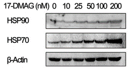
|
28915605 |
| Growth inhibition assay | Cell proliferation |
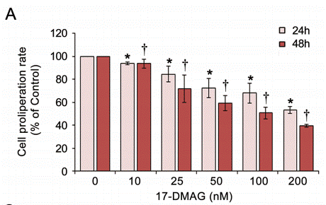
|
28915605 |
Informatie over klinische proeven
(gegevens van https://clinicaltrials.gov, bijgewerkt op 2024-05-22)
| NCT-nummer | Werving | Aandoeningen | Sponsor/medewerkers | Startdatum | Fasen |
|---|---|---|---|---|---|
| NCT00780000 | Terminated | Breast Cancer |
Bristol-Myers Squibb |
April 2008 | Phase 2 |
| NCT00248521 | Unknown status | Unspecified Adult Solid Tumor Protocol Specific |
Institute of Cancer Research United Kingdom|National Cancer Institute (NCI) |
October 2005 | Phase 1 |
Technische ondersteuning
Tel: +1-832-582-8158 Ext:3
Als u nog andere vragen heeft, laat dan een bericht achter.
Producten zijn uitsluitend voor onderzoeksdoeleinden. Niet voor menselijk gebruik. Wij verkopen niet aan patiënten.
©Copyright 2013 Selleck Chemicals. Alle rechten voorbehouden.