alleen voor onderzoeksdoeleinden
NSC 74859 (S3I-201) STAT3-remmer
Cat.nr.S1155
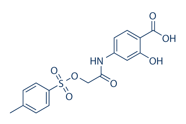
Chemische structuur
Molecuulgewicht: 365.36
Kwaliteitscontrole
| Gerelateerde doelwitten | EGFR JAK Pim |
|---|---|
| Overig STAT Inhibitoren | Napabucasin (BBI608) Stattic Cryptotanshinone (Tanshinone C) C188-9 (TTI-101) SH-4-54 BP-1-102 AS1517499 Nifuroxazide HO-3867 Homoharringtonine (HHT) |
Celkweek, behandeling & werkzame concentratie
| Cellijnen | Assaytype | Concentratie | Incubatietijd | Formulering | Activiteitsbeschrijving | PMID |
|---|---|---|---|---|---|---|
| U87 | Growth Inhibition Assay | 72 h | IC50=55.1 μM | 20072652 | ||
| U373 | Growth Inhibition Assay | 72 h | IC50=52.5 μM | 20072652 | ||
| HPAC | Growth Inhibition Assay | 72 h | IC50>100 μM | 20072652 | ||
| PANC-1 | Growth Inhibition Assay | 72 h | IC50>100 μM | 20072652 | ||
| SK-BR-3 | Growth Inhibition Assay | 72 h | IC50>100 μM | 20072652 | ||
| U-373 MG | Cytotoxicity Assay | 3/10 μM | 24 h | reduces FN-γ-induced cell neurotoxicity | 20888416 | |
| MDA-MB-231 | Growth Inhibition Assay | 72 h | IC50>100 μM | 20072652 | ||
| HUVEC | Function Assay | 0.5-20 μM | 24 h | DMSO | suppresses the hypoxia-induced accumulation of HIF-1α | 21523559 |
| Huh7 | Growth Inhibition Assay | 100 nM | 48 h | DMSO | inhibits the IL-6 stimulation promoted cell proliferation | 23364389 |
| PLC/PRF/5 | Growth Inhibition Assay | 100 nM | 48 h | DMSO | inhibits the IL-6 stimulation promoted cell proliferation | 23364389 |
| H460 | Function Assay | 50/100 μM | 48 h | inhibits the Stat3C increased miR-92a expression | 23820254 | |
| H1299 | Function Assay | 50/100 μM | 48 h | suppresses miR-92a expression dose-dependently | 23820254 | |
| T-cell | Growth Inhibition Assay | IC50=50 μM | 24068731 | |||
| U373 | Growth Inhibition Assay | 125 μM | 24 h | DMSO | disrupts STAT3 signaling and proliferation | 24070820 |
| HUT-102 | Apoptosis Assay | 75-300 μM | 24/48 h | suppresses cell proliferation in a dose-dependent manner and induces cell apoptosis | 24090995 | |
| MT-2 | Apoptosis Assay | 75-300 μM | 24/48 h | suppresses cell proliferation in a dose-dependent manner and induces cell apoptosis | 24090995 | |
| H460 | Apoptosis Assay | 100 nM | 24 h | enhances cell death co-treated with LY294002 | 24472538 | |
| A459 | Apoptosis Assay | 100 nM | 24 h | induces cell apoptosis co-treated with BEZ235 | 24472538 | |
| H460 | Apoptosis Assay | 100 nM | 24 h | induces cell apoptosis co-treated with BEZ235 | 24472538 | |
| GC | Growth Inhibition Assay | 50-125 μM | 72 h | attenuates the cell growth in a dose-dependent manner | 25774503 | |
| GH3 | Growth Inhibition Assay | 50-125 μM | 72 h | attenuates the cell growth in a dose-dependent manner | 25774503 | |
| BT474R | Function Assay | 50 μM | 10-60 d | inhibits STAT3 activity | 25327561 | |
| NCI-N87R | Function Assay | 50 μM | 10-60 d | inhibits STAT3 activity | 25327561 | |
| MDA-MB-468 | Function assay | 100 uM | 24 hrs | Inhibition of Stat3 activation in human MDA-MB-468 cells at 100 uM after 24 hrs | 17463090 | |
| MDA-MB-435 | Function assay | 100 uM | 24 hrs | Inhibition of Stat3 activation in human MDA-MB-435 cells at 100 uM after 24 hrs | 17463090 | |
| MDA-MB-231 | Function assay | 100 uM | 24 hrs | Inhibition of Stat3 activation in human MDA-MB-231 cells at 100 uM after 24 hrs | 17463090 | |
| NIH3T3 | Function assay | 100 uM | 24 hrs | Reduction of pTyr-705 Stat3 level in v-Src expressing mouse NIH3T3 cells at 100 uM after 24 hrs | 17463090 | |
| NIH3T3 | Growth inhibition assay | 100 uM | 4 days | Growth inhibition of mouse NIH3T3 cells expressing v-Src at 100 uM after 4 days by trypan blue exclusion assay | 17463090 | |
| MDA-MB-435 | Growth inhibition assay | 100 uM | 4 days | Growth inhibition of human MDA-MB-435 cells expressing v-Src at 100 uM after 4 days by trypan blue exclusion assay | 17463090 | |
| MDA-MB-231 | Growth inhibition assay | 100 uM | 4 days | Growth inhibition of human MDA-MB-231 cells expressing v-Src at 100 uM after 4 days by trypan blue exclusion assay | 17463090 | |
| MDA-MB-468 | Growth inhibition assay | 100 uM | 4 days | Growth inhibition of human MDA-MB-468 cells expressing v-Src at 100 uM after 4 days by trypan blue exclusion assay | 17463090 | |
| NIH3T3 | Growth inhibition assay | 100 uM | Growth inhibition of mouse NIH3T3 cells expressing v-Ras at 100 uM for every 3 days by soft-agar colony-formation assay | 17463090 | ||
| MDA-MB-435 | Apoptosis assay | 30 to 100 uM | 48 hrs | Induction of apoptosis in human MDA-MB-435 cells expressing active Stat3 at 30 to 100 uM after 48 hrs | 17463090 | |
| MDA-MB-231 | Apoptosis assay | 100 uM | 24 hrs | Reduction of apoptosis in Stat3 transfected human MDA-MB-231 cells at 100 uM after 24 hrs | 17463090 | |
| MDA-MB-231 | Function assay | 100 uM | 48 hrs | Reduction of cyclin D1 gene expression in human MDA-MB-231 cells at 100 uM after 48 hrs | 17463090 | |
| MDA-MB-231 | Apoptosis assay | 100 uM | 24 hrs | Induction of apoptosis in Stat3 SH2 domain transfected human MDA-MB-231 cells at 100 uM after 24 hrs | 17463090 | |
| MDA-MB-231 | Apoptosis assay | 100 uM | 24 hrs | Induction of apoptosis in Stat3C transfected human MDA-MB-231 cells at 100 uM after 24 hrs | 17463090 | |
| NIH3T3 | Function assay | 100 uM | 48 hrs | Reduction of cyclin D1 gene expression in v-Src transfected mouse NIH3T3 cells at 100 uM after 48 hrs | 17463090 | |
| NIH3T3 | Function assay | 100 uM | 48 hrs | Reduction of Bcl-xL gene expression in v-Src transfected mouse NIH3T3 cells at 100 uM after 48 hrs | 17463090 | |
| NIH3T3 | Function assay | 100 uM | 48 hrs | Reduction of survivin gene expression in v-Src transfected mouse NIH3T3 cells at 100 uM after 48 hrs | 17463090 | |
| MDA-MB-231 | Function assay | 100 uM | 48 hrs | Reduction of Bcl-xL gene expression in human MDA-MB-231 cells at 100 uM after 48 hrs | 17463090 | |
| MDA-MB-231 | Function assay | 100 uM | 48 hrs | Reduction of survivin gene expression in human MDA-MB-231 cells at 100 uM after 48 hrs | 17463090 | |
| MDA-MB-231 | Antitumor assay | 5 mg/kg | 2 weeks | Antitumor activity against human MDA-MB-231 cells expressing active Stat3 xenografted in mouse at 5 mg/kg, iv for every 3 days for 2 weeks | 17463090 | |
| NIH3T3 | Growth inhibition assay | 100 uM | Growth inhibition of mouse NIH3T3 cells expressing v-Src at 100 uM for every 3 days by soft-agar colony-formation assay | 17463090 | ||
| A673 | qHTS assay | qHTS of pediatric cancer cell lines to identify multiple opportunities for drug repurposing: Primary screen for A673 cells | 29435139 | |||
| SK-N-MC | qHTS assay | qHTS of pediatric cancer cell lines to identify multiple opportunities for drug repurposing: Primary screen for SK-N-MC cells | 29435139 | |||
| NB1643 | qHTS assay | qHTS of pediatric cancer cell lines to identify multiple opportunities for drug repurposing: Primary screen for NB1643 cells | 29435139 | |||
| LAN-5 | qHTS assay | qHTS of pediatric cancer cell lines to identify multiple opportunities for drug repurposing: Primary screen for LAN-5 cells | 29435139 | |||
| Klik om meer experimentele gegevens over cellijnen te bekijken | ||||||
Chemische informatie, opslag en stabiliteit
| Molecuulgewicht | 365.36 | Formule | C16H15NO7S |
Opslag (vanaf de datum van ontvangst) | |
|---|---|---|---|---|---|
| CAS-nr. | 501919-59-1 | SDF downloaden | Opslag van stamoplossingen |
|
|
Oplosbaarheid
|
In vitro |
DMSO
: 73 mg/mL
(199.8 mM)
Water : Insoluble Ethanol : Insoluble |
Molariteitscalculator
|
In vivo |
|||||
In vivo formulatiecalculator (heldere oplossing)
Stap 1: Voer onderstaande informatie in (Aanbevolen: een extra dier om rekening te houden met verlies tijdens het experiment)
Stap 2: Voer de in vivo formulering in (Dit is alleen de calculator, geen formulering. Neem eerst contact met ons op als er geen in vivo formulering is in de sectie oplosbaarheid.)
Berekeningsresultaten:
Werkconcentratie: mg/ml;
Methode voor het bereiden van DMSO-moedervloeistof: mg geneesmiddel vooropgelost in μL DMSO ( Concentratie moedervloeistof mg/mL, Neem eerst contact met ons op als de concentratie de DMSO-oplosbaarheid van de batch van het geneesmiddel overschrijdt. )
Methode voor het bereiden van in vivo formulering: Neem μL DMSO moedervloeistof, voeg daarna toeμL PEG300, mengen en verhelderen, daarna toevoegenμL Tween 80, mengen en verhelderen, daarna toevoegen μL ddH2O, mengen en verhelderen.
Methode voor het bereiden van in vivo formulering: Neem μL DMSO moedervloeistof, voeg daarna toe μL Maïsolie, mengen en verhelderen.
Opmerking: 1. Zorg ervoor dat de vloeistof helder is voordat u het volgende oplosmiddel toevoegt.
2. Zorg ervoor dat u het/de oplosmiddel(en) in de juiste volgorde toevoegt. U moet ervoor zorgen dat de verkregen oplossing, bij de vorige toevoeging, een heldere oplossing is voordat u verdergaat met het toevoegen van het volgende oplosmiddel. Fysieke methoden zoals vortexen, ultrasoon of een warmwaterbad kunnen worden gebruikt om het oplossen te bevorderen.
Werkingsmechanisme
| Kenmerken |
A chemical probe inhibitor of Stat3 activity.
|
|---|---|
| Targets/IC50/Ki |
STAT3
(Cell-free assay) 86 μM
|
| In vitro |
NSC 74859 (S3I-201) remt de groei en induceert apoptose bij voorkeur in tumorcellen die persistent geactiveerde Stat3 bevatten door de vorming van het Stat3·Stat3-complex en de DNA-bindende en transcriptionele activiteiten van Stat3 te remmen. Bovendien remt het ook de expressie van de Stat3-gereguleerde genen die coderen voor cycline D1, Bcl-xL en survivin. Deze verbinding remt de borstcarcinoom MDA-MB-435, MDA-MB-453 en MDA-MB-231 cellijnen met een IC50 van 100 μM. Bovendien zijn de cellen met een verminderde TGF-β-signalering vier keer zo gevoelig voor S3I-201. Een recente studie toont aan dat het het antiproliferatieve effect in HepG2- en Huh-7-cellen versterkt via de STAT3-signaleringsroute. |
| Kinase Assay |
In vitro Stat3 DNA-bindingstest en EMSA-analyse
|
|
Kort gezegd wordt 100 μL biotinyl-e-Ac-EPQpYEEIEL-OH (in 50 mM Tris/150 mM NaCl, pH 7.5) toegevoegd aan elk putje van met streptavidine gecoate 96-wells microtiterplaten en ‘s nachts bij 4 °C geschud. Vervolgens worden de platen gespoeld met PBS/Tween 20 en daarna tweemaal met 200 μL BSA-T-PBS (0.2% BSA/0.1% Tween 20/PBS). Daarna wordt 50 μL Lck-SH2-GST fusie-eiwit (6.4 ng/ml in BSA-T-PBS) toegevoegd aan elk putje van de 96-wells plaat in aanwezigheid en afwezigheid van 50 μL NSC 74859 (S3I-201) (voor finale concentraties van 30 en 100 mM), en de plaat wordt gedurende 4 uur bij kamertemperatuur geschud. Nadat de oplossingen zijn verwijderd, wordt elk putje viermaal gespoeld met BSA-T-PBS (200 μL), en wordt 100 μL polyclonaal konijnen-anti-GST-antilichaam (100 ng/mL in BSA-T-PBS) toegevoegd aan elk putje en ‘s nachts bij 4 °C geïncubeerd. Na wassen met BSA-T-PBS wordt 100 μL van 200 ng/mL BSA-T-PBS mierikswortelperoxidase-geconjugeerd muis-anti-konijnenantilichaam toegevoegd aan elk putje en gedurende 45 minuten bij kamertemperatuur geïncubeerd. Na vier wasstappen met BSA-T-PBS en drie wasstappen met PBS-T, wordt 100 μL peroxidase-substraat toegevoegd aan elk putje en gedurende 5-15 minuten geïncubeerd. De peroxidase-reactie wordt gestopt door 100 μL 1 M zwavelzuuroplossing toe te voegen, en de absorptie wordt afgelezen bij 450 nm met een ELISA-plaatlezer.
|
|
| In vivo |
NSC 74859 (S3I-201) (5 mg/kg, i.v. elke 2 of elke 3 dagen) toont de antitumorale werkzaamheid in muismodellen met menselijke borsttumor xenografts die constitutief actieve Stat3 bevatten. Deze verbindingbehandeling vermindert Varicella-zoster virus (VZV) replicatie op basis van het bioluminescentiesignaal en het aantal positieve huidxenografts vergeleken met met DMSO behandelde muizen door STAT3-fosforylatie te remmen. |
Referenties |
|
Toepassingen
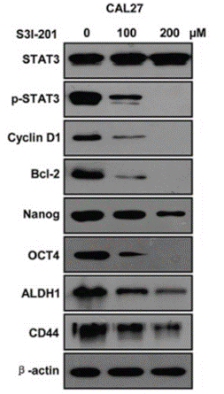
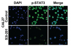
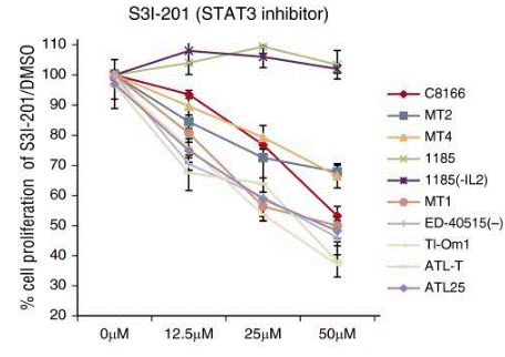
| Methoden | Biomarkers | Afbeeldingen | PMID |
|---|---|---|---|
| Western blot | STAT3 / p-STAT3 / Cyclin D1 / Bcl-2 / Nanog / OCT4 / ALDH1 / CD44 PD-L1 |
|
26556875 |
| Immunofluorescence | p-STAT3 Oct4 / Twist |
|
26556875 |
| Growth inhibition assay | Cell viability |
|
26813676 |
Technische ondersteuning
Tel: +1-832-582-8158 Ext:3
Als u nog andere vragen heeft, laat dan een bericht achter.
Producten zijn uitsluitend voor onderzoeksdoeleinden. Niet voor menselijk gebruik. Wij verkopen niet aan patiënten.
©Copyright 2013 Selleck Chemicals. Alle rechten voorbehouden.