réservé à la recherche
ABT-737 Bcl-2 Inhibiteur
N° Cat.S1002
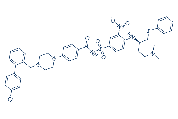
Structure chimique
Poids moléculaire: 813.43
Contrôle qualité
| Cibles apparentées | Caspase PD-1/PD-L1 Ferroptosis p53 Apoptosis related Synthetic Lethality STAT TNF-alpha Ras KRas |
|---|---|
| Autre Bcl-2 Inhibiteurs | Navitoclax (ABT-263) S63845 Obatoclax Mesylate (GX15-070) A-1331852 A-1210477 TW-37 A-1155463 Dihydrochloride AZD5991 UMI-77 (R)-(-)-Gossypol (AT-101) acetic acid |
Culture cellulaire, traitement et concentration de travail
| Lignées cellulaires | Type dessai | Concentration | Temps dincubation | Formulation | Description de lactivité | PMID |
|---|---|---|---|---|---|---|
| OCI-Ly1 | Cell Viability Assay | 250 nM | 72 h | DMSO | caused 97% loss of viability in cells transfected with BCL6 siRNA | 26657288 |
| KG1a | Cell Viability Assay | 0-10 μM | 24 h | DMSO | IC50=7.68 μM, decreases cell viability in a dose-dependent manner | 26552712 |
| Kasumi-1 | Cell Viability Assay | 0-10 μM | 24 h | DMSO | IC50=4.87 μM, decreases cell viability in a dose-dependent manner | 26552712 |
| KG1a | Apoptosis Assay | 0-10 μM | 24 h | DMSO | induces cell apoptosis in a dose-dependent manner | 26552712 |
| Kasumi-1 | Apoptosis Assay | 0-10 μM | 24 h | DMSO | induces cell apoptosis in a dose-dependent manner | 26552712 |
| MC-3 | Growth Inhibition Assay | 5/10/20 μM | 24 h | DMSO | inhibits cell growth in a dose-dependent manner | 26447615 |
| HN22 | Growth Inhibition Assay | 2.5/7.5/22.5 μM | 24 h | DMSO | inhibits cell growth in a dose-dependent manner | 26447615 |
| MC-3 | Apoptosis Assay | 5/10/20 μM | 24 h | DMSO | induces caspase-mediated apoptosis | 26447615 |
| HN22 | Apoptosis Assay | 2.5/7.5/22.5 μM | 24 h | DMSO | induces caspase-mediated apoptosis | 26447615 |
| MOLT-4 | Growth Inhibition Assay | 10-5000 nM | 72 h | DMSO | IC50=0.198 μM | 26392332 |
| RS4;11 | Growth Inhibition Assay | 10-5000 nM | 72 h | DMSO | IC50=0.002 μM | 26392332 |
| JURKAT | Growth Inhibition Assay | 10-5000 nM | 72 h | DMSO | IC50=66 μM | 26392332 |
| CEM R | Growth Inhibition Assay | 10-5000 nM | 72 h | DMSO | IC50=5.4 μM | 26392332 |
| CEM S | Growth Inhibition Assay | 10-5000 nM | 72 h | DMSO | IC50=12.1 μM | 26392332 |
| MOLT-4 | Apoptosis Assay | 10-1000 nM | 24 h | DMSO | causes the cleavage of Bcl-2 and the downregulation of Bcl-xL and Mcl-1 | 26392332 |
| CEM S | Apoptosis Assay | 10-1000 nM | 24 h | DMSO | causes the cleavage of Bcl-2 and the downregulation of Bcl-xL and Mcl-1 | 26392332 |
| JURKAT | Growth Inhibition Assay | 100-1000 nM | 48 h | DMSO | IC50=955±9.3 nM | 26172269 |
| LOUCY | Growth Inhibition Assay | 100-1000 nM | 48 h | DMSO | IC50=32.8±10.9 nM | 26172269 |
| WM-115 | Cell Viability Assay | 100 nM | 72 h | enhances curcumin-induced anti-survival | 26116776 | |
| B16 | Cell Viability Assay | 100 nM | 72 h | enhances curcumin-induced anti-survival | 26116776 | |
| HL-60 | Growth Inhibition Assay | 72 h | IC50 = 10.7 nM | 26045609 | ||
| MOLM-13 | Growth Inhibition Assay | 72 h | IC50 = 27.9 nM | 26045609 | ||
| OCI-AML3 | Growth Inhibition Assay | 72 h | IC50 = 1950 nM | 26045609 | ||
| BCWM.1 | Apoptosis Assay | 0-1.6 μM | 24 h | induces cell apoptosis | 25893290 | |
| MWCL-1 | Apoptosis Assay | 0-1.6 μM | 24 h | induces cell apoptosis | 25893290 | |
| MM.1s | Apoptosis Assay | 0-1.6 μM | 24 h | induces cell apoptosis | 25893290 | |
| HCT116 | Function Assay | 3/10 μM | 12 h | DMSO | induces a dose-dependent increase in LC3B-II conversion and SQSTM1 degradation | 25715028 |
| HCT116 BAX BAK1 DKO | Function Assay | 3/10 μM | 12 h | DMSO | induces a dose-dependent increase in LC3B-II conversion and SQSTM1 degradation | 25715028 |
| HCT116 | Function Assay | 10 μM | 12 h | DMSO | increases GFP-LC3B puncta | 25715028 |
| HCT116 BAX BAK1 DKO | Function Assay | 10 μM | 12 h | DMSO | increases GFP-LC3B puncta | 25715028 |
| HCT116 | Autophagy Assay | 10 μM | 12 h | DMSO | induces a complete autophagic response | 25715028 |
| HCT116 BAX BAK1 DKO | Autophagy Assay | 10 μM | 12 h | DMSO | induces a complete autophagic response | 25715028 |
| U937 | Apoptosis Assay | 0.125-2 μM | 24 h | enhances DHA/X-11-induced apoptosis | 25714024 | |
| U937 | Apoptosis Assay | 0.5 μM | 24 h | enhances cleavage of PARP and caspase-3 as well as Noxa level | 25714024 | |
| HL-60 AAA-Bcl-2 | Apoptosis Assay | 0-5 μM | 48 h | IC50=0.87 μm,induces cell apoptosis in a dose-dependent manner | 25711460 | |
| HL-60 EEE-Bcl-2 | Apoptosis Assay | 0-5 μM | 48 h | IC50=5 μm, induces cell apoptosis in a dose-dependent manner | 25711460 | |
| U87 | Function Assay | 50 μM | 24 h | reduces the mRNA expression levels of MMP-2, MMP-14 and Bcl-2 | 25667663 | |
| K562 | Cell Viability Assay | 1-10 μM | 48 h | DMSO | IC50=26.7 μM | 25596561 |
| K562/Mcl -1-IRESBim | Growth Inhibition Assay | IC50=9.3 μM | 25535900 | |||
| K562/Bcl- 2-IRESBim | Growth Inhibition Assay | IC50=0.35 μM | 25535900 | |||
| Jurkat | Growth Inhibition Assay | IC50=0.66 μM | 25535900 | |||
| JurkatΔBak | Growth Inhibition Assay | IC50>50 μM | 25535900 | |||
| HL60/VCR | Growth Inhibition Assay | IC50>100 μM | 25535900 | |||
| Kasumi-1 | Growth Inhibition Assay | IC50=0.01 μM | 25535900 | |||
| Kasumi-1/ABT | Growth Inhibition Assay | IC50=0.51 μM | 25535900 | |||
| THP-1 | Growth Inhibition Assay | IC50=1.27 μM | 25535900 | |||
| U937 | Growth Inhibition Assay | IC50=5.29 μM | 25535900 | |||
| C1498 | Growth Inhibition Assay | IC50=6.13 μM | 25535900 | |||
| RPMI 8226 | Growth Inhibition Assay | IC50=0.25 μM | 25535900 | |||
| MM.1S | Growth Inhibition Assay | IC50=0.40 μM | 25535900 | |||
| NCI-H929 | Growth Inhibition Assay | IC50=15.21 μM | 25535900 | |||
| U266 | Growth Inhibition Assay | IC50=0.68 μM | 25535900 | |||
| MCF-7 | Cell Viability Assay | 5 μM | 48 h | DMSO | enhances the sensitivity to or radiation | 25409124 |
| MCF-7 | Apoptosis Assay | 5 μM | 4/24/48 h | DMSO | increases the cleaved PARP | 25409124 |
| MCF-7 | Function Assay | 5 μM | 24 h | DMSO | enhances thelevel of Mcl-1 expression | 25409124 |
| MDA-MB 231 | Function Assay | 5 μM | 24 h | DMSO | enhances thelevel of Mcl-1 expression | 25409124 |
| ZR-75-1 | Function Assay | 5 μM | 24 h | DMSO | enhances thelevel of Mcl-1 expression | 25409124 |
| A549 | Cell Viability Assay | 0-20 μM | 72 h | DMSO | decreases the cell survival in a dose-dependent manner combined with aspirin | 25388762 |
| H1299 | Cell Viability Assay | 0-20 μM | 72 h | DMSO | decreases the cell survival in a dose-dependent manner combined with aspirin | 25388762 |
| HO-8910 | Cell Viability Assay | 0-20 μM | 72 h | DMSO | decreases the cell survival in a dose-dependent manner combined with aspirin | 25388762 |
| HT-29 | Cell Viability Assay | 0-20 μM | 72 h | DMSO | decreases the cell survival in a dose-dependent manner combined with aspirin | 25388762 |
| HCT-116 | Cell Viability Assay | 0-20 μM | 72 h | DMSO | decreases the cell survival in a dose-dependent manner combined with aspirin | 25388762 |
| A549 | Apoptosis Assay | 20 μM | 48 h | DMSO | induces apoptosis significantly combined with aspirin | 25388762 |
| H1299 | Apoptosis Assay | 20 μM | 48 h | DMSO | induces apoptosis significantly combined with aspirin | 25388762 |
| Sc-1 | Cell Viability Assay | 0.0001-1 μM | 96 h | decreases the cell viability in a dose-dependent manner | 25373508 | |
| OcI-LY18 | Cell Viability Assay | 0.0001-1 μM | 96 h | decreases the cell viability in a dose-dependent manner | 25373508 | |
| RL | Cell Viability Assay | 0.0001-1 μM | 96 h | decreases the cell viability in a dose-dependent manner | 25373508 | |
| RKO | Cell Viability Assay | 0-10 μM | 24 h | DMSO | IC50> 25 µM | 25304383 |
| Caco-2 | Cell Viability Assay | 0-10 μM | 24 h | DMSO | IC50=19.7 µM | 25304383 |
| DLD1 | Cell Viability Assay | 0-10 μM | 24 h | DMSO | IC50=18.78 µM | 25304383 |
| LS411N | Cell Viability Assay | 0-10 μM | 24 h | DMSO | IC50=11.47 µM | 25304383 |
| SW620 | Cell Viability Assay | 0-10 μM | 24 h | DMSO | IC50=12.24 µM | 25304383 |
| HCT116 | Cell Viability Assay | 0-10 μM | 24 h | DMSO | IC50=20.49 µM | 25304383 |
| HaCaT | Cell Viability Assay | 0.1/1/10 μM | 24 h | DMSO | decreases cell viability in a dose-dependent manner | 25210795 |
| A5-RT3 | Cell Viability Assay | 0.1/1/10 μM | 24 h | DMSO | decreases cell viability in a dose-dependent manner | 25210795 |
| HaCaT | Function Assay | 10 μM | 24/48 h | DMSO | induces MMP and DNA fragmentation | 25210795 |
| A5-RT3 | Function Assay | 10 μM | 24/48 h | DMSO | induces MMP and DNA fragmentation | 25210795 |
| A5-RT3 | Function Assay | 5 μM | 6 h | DMSO | induces the release of mitochondrial proteins and reduces clonogenic survival in a caspase-independent manner | 25210795 |
| U266 | Function Assay | 500/750 nM | 24/48 h | DMSO | downregulates Bim, principally the EL isoform | 25208888 |
| RPMI8226 | Function Assay | 500/750 nM | 24/48 h | DMSO | downregulates Bim, principally the EL isoform | 25208888 |
| MM.1S | Function Assay | 500/750 nM | 24/48 h | DMSO | downregulates Bim, principally the EL isoform | 25208888 |
| Clone A | Growth Inhibition Assay | 0.2–60 μM | 72 h | DMSO | IC50=7.5 μM | 25208882 |
| CX-1 | Growth Inhibition Assay | 0.2–60 μM | 72 h | DMSO | IC50=1.8 μM | 25208882 |
| LS174T | Growth Inhibition Assay | 0.2–60 μM | 72 h | DMSO | IC50=18.3 μM | 25208882 |
| HT29 | Apoptosis Assay | 1/5/10 μM | 48 h | causes cell death in a dose-dependent manner | 25192188 | |
| SW480 | Apoptosis Assay | 1/5/10 μM | 48 h | causes cell death in a dose-dependent manner | 25192188 | |
| Colo205 | Apoptosis Assay | 1/5/10 μM | 48 h | causes cell death in a dose-dependent manner | 25192188 | |
| Caco2 | Apoptosis Assay | 1/5/10 μM | 48 h | causes cell death in a dose-dependent manner | 25192188 | |
| PCI-13 | Growth Inhibition Assay | 72 h | DMSO | GI50=15 ± 1.8 μM | 25139387 | |
| PCI-15B | Growth Inhibition Assay | 72 h | DMSO | GI50=11 ± 4.5 μM | 25139387 | |
| UM-SCC22B | Growth Inhibition Assay | 72 h | DMSO | GI50=19 ± 2.9 μM | 25139387 | |
| UM-SCC47 | Growth Inhibition Assay | 72 h | DMSO | GI50=19 ± 12.3 μM | 25139387 | |
| 93-VU-147T | Growth Inhibition Assay | 72 h | DMSO | GI50=4.3 ± 3.5 μM | 25139387 | |
| UD-SCC2 | Growth Inhibition Assay | 72 h | DMSO | GI50=28 ± 2.9 μM | 25139387 | |
| UPCI:SCC90 | Growth Inhibition Assay | 72 h | DMSO | GI50=6.6 ± 1.5 μM | 25139387 | |
| RPMI-8226 | Cell Viability Assay | 125/250/500 nM | 48h | DMSO | decreases cell viability in a dose-dependent manner | 25008202 |
| OPM-2 | Cell Viability Assay | 125/250/500 nM | 48h | DMSO | decreases cell viability in a dose-dependent manner | 25008202 |
| RPMI-8226 | Apoptosis Assay | 125/250/500 nM | 48h | DMSO | induces cell apoptosis in a dose-dependent manner | 25008202 |
| OPM-2 | Apoptosis Assay | 125/250/500 nM | 48h | DMSO | induces cell apoptosis in a dose-dependent manner | 25008202 |
| COG-LL-319 | Function Assay | 100 nM | 1/3/6 h | DMSO | induces caspase-dependent Mcl-1 cleavage | 24951472 |
| RS4;11 | Function Assay | 100 nM | 1/3/6 h | DMSO | induces caspase-dependent Mcl-1 cleavage | 24951472 |
| FL5.12 | Function assay | Reversal of cytokine withdrawal protection in IL3 dependent Bcl2 overexpressing mouse FL5.12 cells in presence of bovine gelatin, EC50 = 0.008 μM. | 17256834 | |||
| DoHH2 | Growth inhibition assay | Inhibition of cell growth in human DoHH2 cells overexpressing Bcl2 in presence of 3% FBS, EC50 = 0.0083 μM. | 17256834 | |||
| RS11380 | Growth inhibition assay | Inhibition of cell growth in human RS11380 cells overexpressing Bcl2 in presence of 3% FBS, EC50 = 0.014 μM. | 17256834 | |||
| FL5.12 | Function assay | Reversal of cytokine withdrawal protection in IL3 dependent Bcl-xL overexpressing mouse FL5.12 cells in presence of bovine gelatin, EC50 = 0.03 μM. | 17256834 | |||
| FL5.12 | Function assay | Reversal of cytokine withdrawal protection in IL3 dependent Bcl2 overexpressing mouse FL5.12 cells in presence of 3% FBS, EC50 = 0.05 μM. | 17256834 | |||
| DoHH2 | Growth inhibition assay | Inhibition of cell growth in human DoHH2 cells overexpressing Bcl2 in presence of 10% HS, EC50 = 0.13 μM. | 17256834 | |||
| RS11380 | Growth inhibition assay | Inhibition of cell growth in human RS11380 cells overexpressing Bcl2 in presence of 10% HS, EC50 = 0.15 μM. | 17256834 | |||
| SUDHL4 | Growth inhibition assay | Inhibition of cell growth in human SUDHL4 cells overexpressing Bcl2 in presence of 3% FBS, EC50 = 0.22 μM. | 17256834 | |||
| FL5.12 | Function assay | Reversal of cytokine withdrawal protection in IL3 dependent Bcl-xL overexpressing mouse FL5.12 cells in presence of 3% FBS, EC50 = 0.22 μM. | 17256834 | |||
| SUDHL4 | Growth inhibition assay | Inhibition of cell growth in human SUDHL4 cells overexpressing Bcl2 in presence of 10% HS, EC50 = 0.85 μM. | 17256834 | |||
| FL5.12 | Cytotoxicity assay | 24 hrs | Cytotoxicity against IL3-dependent mouse FL5.12 cells overexpressing human Bcl2 assessed as cell viability after 24 hrs by MTS assay in absence of serum, EC50 = 0.0077 μM. | 18841882 | ||
| FL5.12 | Cytotoxicity assay | 24 hrs | Cytotoxicity against IL3-dependent mouse FL5.12 cells overexpressing human Bcl-XL assessed as cell viability after 24 hrs by MTS assay in absence of serum, EC50 = 0.03 μM. | 18841882 | ||
| NCI-H146 | Cytotoxicity assay | 48 hrs | Cytotoxicity against human NCI-H146 cells assessed as cell viability after 48 hrs in presence of 10% human serum, EC50 = 0.087 μM. | 18841882 | ||
| CLL | Apoptosis assay | Induction of apoptosis in human CLL cells, EC50 = 0.0045 μM. | 20925433 | |||
| MEF | Cytotoxicity assay | 24 hrs | Cytotoxicity against mouse mcl-1 deficient MEF cells after 24 hrs by Cell titer glo assay in presence of 1% serum, EC50 = 0.00203 μM. | 21366295 | ||
| MEF | Cytotoxicity assay | 24 hrs | Cytotoxicity against mouse mcl-1 deficient MEF cells after 24 hrs by Cell titer glo assay in presence of 10% serum, EC50 = 0.051 μM. | 21366295 | ||
| MEF | Cytotoxicity assay | 24 hrs | Cytotoxicity against mouse mcl-1 deficient MEF cells after 24 hrs by Cell titer glo assay in presence of 10% fetal bovine serum, EC50 = 0.051 μM. | 21366295 | ||
| RS4:11 | Antiproliferative assay | Antiproliferative activity against human RS4:11 cells in presence of 10% human serum, EC50 = 0.024 μM. | 28926247 | |||
| MOLT4 | Antiproliferative assay | Antiproliferative activity against human MOLT4 cells in presence of 10% human serum, EC50 = 0.622 μM. | 28926247 | |||
| Jurkat | Cytotoxicity assay | 48 hrs | Cytotoxicity against human Jurkat cells after 48 hrs by cell titer-blue assay, IC50 = 1.38 μM. | 19743858 | ||
| HCT116 | Cytotoxicity assay | 48 hrs | Cytotoxicity against human HCT116 cells after 48 hrs by cell titer-blue assay, IC50 = 4.06 μM. | 19743858 | ||
| SU-8686 | Cytotoxicity assay | 48 hrs | Cytotoxicity against human SU-8686 cells after 48 hrs by cell titer-blue assay, IC50 = 4.24 μM. | 19743858 | ||
| H460 | Cytotoxicity assay | 48 hrs | Cytotoxicity against human H460 cells after 48 hrs by cell titer-blue assay, IC50 = 8.03 μM. | 19743858 | ||
| Hepa-1c1c7 | Cytotoxicity assay | 48 hrs | Cytotoxicity against mouse Hepa-1c1c7 cells after 48 hrs by cell titer-blue assay, IC50 = 8.68 μM. | 19743858 | ||
| MCF7 | Cytotoxicity assay | 48 hrs | Cytotoxicity against human MCF7 cells after 48 hrs by cell titer-blue assay, IC50 = 21.26 μM. | 19743858 | ||
| DU145 | Cytotoxicity assay | 48 hrs | Cytotoxicity against human DU145 cells after 48 hrs by cell titer-blue assay, IC50 = 27.6 μM. | 19743858 | ||
| HCT116 | Cytotoxicity assay | 72 hrs | Cytotoxicity against human HCT116 cells expressing Bcl-xL, Bcl-2 and Mcl-1 after 72 hrs by MTT assay, IC50 = 47.7 μM. | 22172701 | ||
| BL21 (DE3) | Function assay | 2 hrs | Binding affinity to N-terminus 6X His-tagged human Bcl2 expressed in Escherichia coli BL21 (DE3) cells after 2 hrs by fluorescence polarization assay, IC50 = 0.002 μM. | 22448988 | ||
| BL21 (DE3) | Function assay | 2 hrs | Binding affinity to N-terminus 8X His-tagged human Bcl-xL expressed in Escherichia coli BL21 (DE3) cells after 2 hrs by fluorescence polarization assay, IC50 = 0.006 μM. | 22448988 | ||
| NCI-H146 | Growth inhibition assay | 4 days | Growth inhibition of human NCI-H146 cells after 4 days by WST8 assay, IC50 = 0.097 μM. | 22448988 | ||
| NCI-H1417 | Growth inhibition assay | 4 days | Growth inhibition of human NCI-H1417 cells after 4 days by WST8 assay, IC50 = 0.13 μM. | 22448988 | ||
| CCRF-CEM | Cytotoxicity assay | 48 hrs | Cytotoxicity against human CCRF-CEM cells assessed as cell viability after 48 hrs by celltiter-blue assay, IC50 = 0.74 μM. | 22582991 | ||
| HL60 | Cytotoxicity assay | 48 hrs | Cytotoxicity against human HL60 cells assessed as cell viability after 48 hrs by celltiter-blue assay, IC50 = 0.76 μM. | 22582991 | ||
| K562 | Cytotoxicity assay | 48 hrs | Cytotoxicity against human K562 cells assessed as cell viability after 48 hrs by celltiter-blue assay, IC50 = 34.7 μM. | 22582991 | ||
| NCI-H146 | Antiproliferative assay | 4 days | Antiproliferative activity against human NCI-H146 cells after 4 days by WST8 assay, IC50 = 0.037 μM. | 22747598 | ||
| NCI-H1963 | Antiproliferative assay | 4 days | Antiproliferative activity against human NCI-H1963 cells after 4 days by WST8 assay, IC50 = 0.059 μM. | 22747598 | ||
| NCI-H1417 | Antiproliferative assay | 4 days | Antiproliferative activity against human NCI-H1417 cells after 4 days by WST8 assay, IC50 = 0.412 μM. | 22747598 | ||
| RS4:11 | Apoptosis assay | 48 hrs | Induction of apoptosis in Bcl2 dependent human RS4:11 cells after 48 hrs by Annexin V staining based flow cytometry, IC50 = 0.27 μM. | 23314054 | ||
| K562 | Apoptosis assay | 48 hrs | Induction of apoptosis in Mcl1 dependent human K562 cells after 48 hrs by Annexin V staining based flow cytometry, IC50 = 16.4 μM. | 23314054 | ||
| NCI-H1963 | Cytotoxicity assay | 4 days | Cytotoxicity against human NCI-H1963 cells assessed as growth inhibition after 4 days by WST assay, IC50 = 0.054 μM. | 23448298 | ||
| NCI-H187 | Cytotoxicity assay | 4 days | Cytotoxicity against human NCI-H187 cells assessed as growth inhibition after 4 days by WST assay, IC50 = 0.1377 μM. | 23448298 | ||
| NCI-H1417 | Cytotoxicity assay | 4 days | Cytotoxicity against human NCI-H1417 cells assessed as growth inhibition after 4 days by WST assay, IC50 = 0.1734 μM. | 23448298 | ||
| HL60 | Growth inhibition assay | 72 hrs | Growth inhibition of human HL60 cells after 72 hrs by MTT assay, IC50 = 0.97 μM. | 27712939 | ||
| MCF7 | Growth inhibition assay | 72 hrs | Growth inhibition of human MCF7 cells after 72 hrs by MTT assay, IC50 = 25.33 μM. | 27712939 | ||
| U266 | Growth inhibition assay | 72 hrs | Growth inhibition of human U266 cells after 72 hrs by MTT assay, IC50 = 27.35 μM. | 27712939 | ||
| SKOV3 | Growth inhibition assay | 72 hrs | Growth inhibition of human SKOV3 cells after 72 hrs by MTT assay, IC50 = 46.59 μM. | 27712939 | ||
| RS4:11 | Cytotoxicity assay | 24 hrs | Cytotoxicity against human RS4:11 cells assessed as reduction in cell viability after 24 hrs by MTT assay, IC50 = 0.33 μM. | 29453135 | ||
| Remb1 | Cytotoxicity assay | 24 hrs | Cytotoxicity against human Remb1 cells assessed as reduction in cell viability after 24 hrs by MTT assay, IC50 = 1.4 μM. | 29453135 | ||
| BL21 | Function assay | 10 uM | 3 hrs | Displacement of BODIPY-Bak conjugated peptide from GST-tagged human Bcl-2-like protein 1 G196A mutant expressed in Escherichia coli BL21 cells at 10 uM after 3 hrs by fluorescence polarization competition assay, Ki = 0.001 μM. | 21807512 | |
| BL21 | Function assay | 10 uM | 3 hrs | Displacement of BODIPY-Bak conjugated peptide from GST-tagged human Bcl-2-like protein 1 A93V mutant expressed in Escherichia coli BL21 cells at 10 uM after 3 hrs by fluorescence polarization competition assay, Ki = 0.0014 μM. | 21807512 | |
| BL21 | Function assay | 10 uM | 3 hrs | Displacement of BODIPY-Bak conjugated peptide from GST-tagged human Bcl-2-like protein 1 Y195F mutant expressed in Escherichia coli BL21 cells at 10 uM after 3 hrs by fluorescence polarization competition assay, Ki = 0.0015 μM. | 21807512 | |
| BL21 | Function assay | 10 uM | 3 hrs | Displacement of BODIPY-Bak conjugated peptide from GST-tagged human Wild type Bcl-2-like protein 1 expressed in Escherichia coli BL21 cells at 10 uM after 3 hrs by fluorescence polarization competition assay, Ki = 0.0034 μM. | 21807512 | |
| BL21 | Function assay | 10 uM | 3 hrs | Displacement of BODIPY-Bak conjugated peptide from GST-tagged human Bcl-2-like protein 1 E129H mutant expressed in Escherichia coli BL21 cells at 10 uM after 3 hrs by fluorescence polarization competition assay, Ki = 0.0045 μM. | 21807512 | |
| BL21 | Function assay | 10 uM | 3 hrs | Displacement of BODIPY-Bak conjugated peptide from GST-tagged human Bcl-2-like protein 1 E96G mutant expressed in Escherichia coli BL21 cells at 10 uM after 3 hrs by fluorescence polarization competition assay, Ki = 0.0058 μM. | 21807512 | |
| BL21 | Function assay | 10 uM | 3 hrs | Displacement of BODIPY-Bak conjugated peptide from GST-tagged human Bcl-2-like protein 1 A142Gdelta136T mutant expressed in Escherichia coli BL21 cells at 10 uM after 3 hrs by fluorescence polarization competition assay, Ki = 0.01 μM. | 21807512 | |
| BL21 | Function assay | 10 uM | 3 hrs | Displacement of BODIPY-Bak conjugated peptide from GST-tagged human Bcl-2-like protein 1 A142G mutant expressed in Escherichia coli BL21 cells at 10 uM after 3 hrs by fluorescence polarization competition assay, Ki = 0.021 μM. | 21807512 | |
| BL21 | Function assay | 10 uM | 3 hrs | Displacement of BODIPY-Bak conjugated peptide from GST-tagged human Bcl-2-like protein 1 L130V mutant expressed in Escherichia coli BL21 cells at 10 uM after 3 hrs by fluorescence polarization competition assay, Ki = 0.058 μM. | 21807512 | |
| BL21 | Function assay | 10 uM | 3 hrs | Displacement of BODIPY-Bak conjugated peptide from GST-tagged human Bcl-2-like protein 1 delta136T mutant expressed in Escherichia coli BL21 cells at 10 uM after 3 hrs by fluorescence polarization competition assay, Ki = 0.06 μM. | 21807512 | |
| BL21 | Function assay | 10 uM | 3 hrs | Displacement of BODIPY-Bak conjugated peptide from GST-tagged human Bcl-2-like protein 1 L130A mutant expressed in Escherichia coli BL21 cells at 10 uM after 3 hrs by fluorescence polarization competition assay, Ki = 0.073 μM. | 21807512 | |
| BL21 | Function assay | 10 uM | 3 hrs | Displacement of BODIPY-Bak conjugated peptide from GST-tagged human Bcl-2-like protein 1 R100E mutant expressed in Escherichia coli BL21 cells at 10 uM after 3 hrs by fluorescence polarization competition assay, Ki = 0.16 μM. | 21807512 | |
| BL21 | Function assay | 10 uM | 3 hrs | Displacement of BODIPY-Bak conjugated peptide from GST-tagged human Bcl-2-like protein 1 V141A mutant expressed in Escherichia coli BL21 cells at 10 uM after 3 hrs by fluorescence polarization competition assay, Ki = 0.19 μM. | 21807512 | |
| BL21 | Function assay | 10 uM | 3 hrs | Displacement of BODIPY-Bak conjugated peptide from GST-tagged human Bcl-2-like protein 1 A142T mutant expressed in Escherichia coli BL21 cells at 10 uM after 3 hrs by fluorescence polarization competition assay, Ki = 0.27 μM. | 21807512 | |
| BL21 | Function assay | 10 uM | 3 hrs | Displacement of BODIPY-Bak conjugated peptide from GST-tagged human Bcl-2-like protein 1 L130G mutant expressed in Escherichia coli BL21 cells at 10 uM after 3 hrs by fluorescence polarization competition assay, Ki = 0.29 μM. | 21807512 | |
| BL21 | Function assay | 10 uM | 3 hrs | Displacement of BODIPY-Bak conjugated peptide from GST-tagged human Bcl-2-like protein 1 F97V mutant expressed in Escherichia coli BL21 cells at 10 uM after 3 hrs by fluorescence polarization competition assay, Ki = 0.3 μM. | 21807512 | |
| BL21 | Function assay | 10 uM | 3 hrs | Displacement of BODIPY-Bak conjugated peptide from GST-tagged human Bcl-2-like protein 1 Y101H mutant expressed in Escherichia coli BL21 cells at 10 uM after 3 hrs by fluorescence polarization competition assay, Ki = 0.38 μM. | 21807512 | |
| BL21 (DE3) | Function assay | 2 hrs | Binding affinity to N-terminus 6X His-tagged human Bcl2 expressed in Escherichia coli BL21 (DE3) cells after 2 hrs by fluorescence polarization assay, Ki = 0.0006 μM. | 22448988 | ||
| BL21 (DE3) | Function assay | 2 hrs | Binding affinity to N-terminus 8X His-tagged human Bcl-xL expressed in Escherichia coli BL21 (DE3) cells after 2 hrs by fluorescence polarization assay, Ki = 0.001 μM. | 22448988 | ||
| Toledo | Apoptosis assay | Induction of apoptosis in human Toledo cells, LD50 = 0.06 μM. | 24900652 | |||
| KB | Cytotoxicity assay | 0.5 uM | Cytotoxicity in human siRNA-mediated-MCL1-kncok down KB cells overexpressing BCL2 at 0.5 uM | 18040043 | ||
| Eu-Myc | Apoptosis assay | 1 uM | Induction of apoptosis in mouse Eu-Myc cells overexpressing BCL2 assessed as inhibition of colony formation at 1 uM | 18040043 | ||
| HeLa | Function assay | 1 uM | 12 hrs | Induction of Bcl-xL-mediated apoptosis in doxycyclin-stimulated human HeLa cells overexpressing Noxa at 1 uM after 12 hrs by Hoechst staining | 22386982 | |
| HeLa | Function assay | 10 uM | 16 hrs | Inhibition of Rluc-Bax/eYFP-Bcl-xL interaction expressed in human HeLa cells at 10 uM after 16 hrs by BRET assay | 22425031 | |
| MDA-MB-231 | Function assay | 0.03 to 1 uM | 1 hr | Antagonist activity at recombinant Bcl-XL assessed as restoration of BIM BH3-induced Smac protein release in mitochondria isolated from MDA-MB-231 cells at 0.03 to 1 uM after 1 hr by Western blot analysis | 22747598 | |
| MDA-MB-231 | Function assay | 0.03 to 1 uM | 1 hr | Antagonist activity at recombinant Bcl-XL assessed as restoration of BIM BH3-induced cytochrome c release in mitochondria isolated from MDA-MB-231 cells at 0.03 to 1 uM after 1 hr by Western blot analysis | 22747598 | |
| BP3 | Apoptosis assay | 10 uM | 24 hrs | Induction of apoptosis in human BP3 cells at 10 uM incubated for 24 hrs by Annexin V and propidium iodide staining based FACS method | 23047228 | |
| IM9 | Apoptosis assay | 10 uM | 24 hrs | Induction of apoptosis in human IM9 cells at 10 uM incubated for 24 hrs by Annexin V and propidium iodide staining based FACS method | 23047228 | |
| RS4:11 | Apoptosis assay | 10 uM | 24 hrs | Induction of apoptosis in human RS4:11 cells at 10 uM incubated for 24 hrs by Annexin V and propidium iodide staining based FACS method | 23047228 | |
| HCT116 | Apoptosis assay | 48 hrs | Induction of apoptosis in human HCT116 p53+/+ cells after 48 hrs by Annexin V-FITC staining-based flow cytometric method | 26982372 | ||
| DMS53 | Apoptosis assay | 5 to 10 uM | 12 hrs | Induction of apoptosis in human DMS53 cells harboring p53 mutant assessed as cytochrome c release at 5 to 10 uM after 12 hrs by immunoblotting method | 26982372 | |
| DMS53 | Function assay | 5 to 10 uM | 12 hrs | Inhibition of BCl-2/Bim interaction in human DMS53 cells harboring p53 mutant at 5 to 10 uM after 12 hrs by immunoprecipitation method | 26982372 | |
| DMS53 | Apoptosis assay | 5 to 10 uM | 12 hrs | Induction of apoptosis in human DMS53 cells harboring p53 mutant assessed as PARP cleavage at 5 to 10 uM after 12 hrs by immunoblotting method | 26982372 | |
| DMS53 | Apoptosis assay | 5 to 10 uM | 12 hrs | Induction of apoptosis in human DMS53 cells harboring p53 mutant assessed as caspase-3 cleavage at 5 to 10 uM after 12 hrs by immunoblotting method | 26982372 | |
| DMS53 | Function assay | 5 to 10 uM | 12 hrs | Inhibition of BCl-2/Bax interaction in human DMS53 cells harboring p53 mutant at 5 to 10 uM after 12 hrs by immunoprecipitation method | 26982372 | |
| U-2 OS | qHTS assay | qHTS of pediatric cancer cell lines to identify multiple opportunities for drug repurposing: Primary screen for U-2 OS cells | 29435139 | |||
| A673 | qHTS assay | qHTS of pediatric cancer cell lines to identify multiple opportunities for drug repurposing: Primary screen for A673 cells | 29435139 | |||
| DAOY | qHTS assay | qHTS of pediatric cancer cell lines to identify multiple opportunities for drug repurposing: Primary screen for DAOY cells | 29435139 | |||
| Saos-2 | qHTS assay | qHTS of pediatric cancer cell lines to identify multiple opportunities for drug repurposing: Primary screen for Saos-2 cells | 29435139 | |||
| RD | qHTS assay | qHTS of pediatric cancer cell lines to identify multiple opportunities for drug repurposing: Primary screen for RD cells | 29435139 | |||
| SK-N-SH | qHTS assay | qHTS of pediatric cancer cell lines to identify multiple opportunities for drug repurposing: Primary screen for SK-N-SH cells | 29435139 | |||
| NB1643 | qHTS assay | qHTS of pediatric cancer cell lines to identify multiple opportunities for drug repurposing: Primary screen for NB1643 cells | 29435139 | |||
| OHS-50 | qHTS assay | qHTS of pediatric cancer cell lines to identify multiple opportunities for drug repurposing: Primary screen for OHS-50 cells | 29435139 | |||
| LAN-5 | qHTS assay | qHTS of pediatric cancer cell lines to identify multiple opportunities for drug repurposing: Confirmatory screen for LAN-5 cells | 29435139 | |||
| DAOY | qHTS assay | qHTS of pediatric cancer cell lines to identify multiple opportunities for drug repurposing: Confirmatory screen for DAOY cells | 29435139 | |||
| NB-EBc1 | qHTS assay | qHTS of pediatric cancer cell lines to identify multiple opportunities for drug repurposing: Confirmatory screen for NB-EBc1 cells | 29435139 | |||
| Rh41 | qHTS assay | qHTS of pediatric cancer cell lines to identify multiple opportunities for drug repurposing: Primary screen for Rh41 cells | 29435139 | |||
| A673 | qHTS assay | qHTS of pediatric cancer cell lines to identify multiple opportunities for drug repurposing: Confirmatory screen for A673 cells) | 29435139 | |||
| Rh30 | qHTS assay | qHTS of pediatric cancer cell lines to identify multiple opportunities for drug repurposing: Primary screen for Rh30 cells | 29435139 | |||
| MG 63 (6-TG R) | qHTS assay | qHTS of pediatric cancer cell lines to identify multiple opportunities for drug repurposing: Confirmatory screen for MG 63 (6-TG R) cells | 29435139 | |||
| U-2 OS | qHTS assay | qHTS of pediatric cancer cell lines to identify multiple opportunities for drug repurposing: Confirmatory screen for U-2 OS cells | 29435139 | |||
| Rh41 | qHTS assay | qHTS of pediatric cancer cell lines to identify multiple opportunities for drug repurposing: Confirmatory screen for Rh41 cells | 29435139 | |||
| SJ-GBM2 | qHTS assay | qHTS of pediatric cancer cell lines to identify multiple opportunities for drug repurposing: Primary screen for SJ-GBM2 cells | 29435139 | |||
| SK-N-MC | qHTS assay | qHTS of pediatric cancer cell lines to identify multiple opportunities for drug repurposing: Primary screen for SK-N-MC cells | 29435139 | |||
| NB-EBc1 | qHTS assay | qHTS of pediatric cancer cell lines to identify multiple opportunities for drug repurposing: Primary screen for NB-EBc1 cells | 29435139 | |||
| LAN-5 | qHTS assay | qHTS of pediatric cancer cell lines to identify multiple opportunities for drug repurposing: Primary screen for LAN-5 cells | 29435139 | |||
| Rh18 | qHTS assay | qHTS of pediatric cancer cell lines to identify multiple opportunities for drug repurposing: Primary screen for Rh18 cells | 29435139 | |||
| NB1643 | qHTS assay | qHTS of pediatric cancer cell lines to identify multiple opportunities for drug repurposing: Confirmatory screen for NB1643 cells | 29435139 | |||
| SK-N-MC | qHTS assay | qHTS of pediatric cancer cell lines to identify multiple opportunities for drug repurposing: Confirmatory screen for SK-N-MC cells | 29435139 | |||
| TC32 | qHTS assay | qHTS of pediatric cancer cell lines to identify multiple opportunities for drug repurposing: Confirmatory screen for TC32 cells | 29435139 | |||
| Rh18 | qHTS assay | qHTS of pediatric cancer cell lines to identify multiple opportunities for drug repurposing: Confirmatory screen for Rh18 cells | 29435139 | |||
| Saos-2 | qHTS assay | qHTS of pediatric cancer cell lines to identify multiple opportunities for drug repurposing: Confirmatory screen for Saos-2 cells | 29435139 | |||
| Cliquez pour voir plus de données expérimentales sur les lignées cellulaires | ||||||
Informations chimiques, stockage et stabilité
| Poids moléculaire | 813.43 | Formule | C42H45ClN6O5S2 |
Stockage (À partir de la date de réception) | |
|---|---|---|---|---|---|
| N° CAS | 852808-04-9 | Télécharger le SDF | Stockage des solutions mères |
|
|
| Synonymes | N/A | Smiles | CN(C)CCC(CSC1=CC=CC=C1)NC2=C(C=C(C=C2)S(=O)(=O)NC(=O)C3=CC=C(C=C3)N4CCN(CC4)CC5=CC=CC=C5C6=CC=C(C=C6)Cl)[N+](=O)[O-] | ||
Solubilité
|
In vitro |
DMSO
: 163 mg/mL
(200.38 mM)
Water : Insoluble Ethanol : Insoluble |
Calculateur de molarité
|
In vivo |
|||||
Calculateur de formulation in vivo (Solution claire)
Étape 1 : Entrez les informations ci-dessous (Recommandé : Un animal supplémentaire pour tenir compte des pertes pendant lexpérience)
Étape 2 : Entrez la formulation in vivo (Ceci nest que le calculateur, pas la formulation. Veuillez nous contacter dabord sil ny a pas de formulation in vivo dans la section Solubilité.)
Résultats du calcul :
Concentration de travail : mg/ml;
Méthode de préparation du liquide maître DMSO : mg médicament prédissous dans μL DMSO ( Concentration du liquide maître mg/mL, Veuillez nous contacter dabord si la concentration dépasse la solubilité du DMSO du lot de médicament. )
Méthode de préparation de la formulation in vivo : Prendre μL DMSO liquide maître, ajouter ensuiteμL PEG300, mélanger et clarifier, ajouter ensuiteμL Tween 80, mélanger et clarifier, ajouter ensuite μL ddH2O, mélanger et clarifier.
Méthode de préparation de la formulation in vivo : Prendre μL DMSO liquide maître, ajouter ensuite μL Huile de maïs, mélanger et clarifier.
Remarque : 1. Assurez-vous que le liquide est clair avant dajouter le solvant suivant.
2. Assurez-vous dajouter le(s) solvant(s) dans lordre. Vous devez vous assurer que la solution obtenue lors de lajout précédent est une solution claire avant de procéder à lajout du solvant suivant. Des méthodes physiques telles que le vortex, les ultrasons ou le bain-marie peuvent être utilisées pour faciliter la dissolution.
Mécanisme daction
| Fonctionnalités |
First-generation inhibitor of anti-apoptotic Bcl-2 proteins.
|
|---|---|
| Targets/IC50/Ki |
Bcl-2
(Cell-free assay) 30.3 nM(EC50)
Bcl-xL
(Cell-free assay) 78.7 nM(EC50)
Bcl-w
(Cell-free assay) 197.8 nM(EC50)
Bcl-B
(Cell-free assay) 1.82 μM(EC50)
|
| In vitro |
ABT-737 montre une faible activité envers Bcl-B et aucun effet envers Mcl-1 et BFL-1. Ce composé est sensible aux cellules HL60, KG1 et NB4 avec des IC50 de 50 nM, 80 nM et 80 nM, respectivement. Il induit l'Apoptosis dans les cellules HL60, ce qui est dû à une hétérodimérisation Bcl-2/Bax diminuée et n'a aucun effet sur la distribution du cycle cellulaire. Il induit également la libération de cytochrome c des mitochondries purifiées et favorise les changements conformationnels de Bax qui sont associés à l'Apoptosis. Les cellules résistantes (Hela et MCF-7) peuvent être sensibilisées à ce produit chimique par des approches qui régulent à la baisse, déstabilisent ou inactivent Mcl-1. Il provoque également la libération de cytochrome c dépendante de Bax/BAK uniquement lorsque Mcl-1 a été neutralisé. Ce composé déplace Bim de la poche de liaison BH3 de Bcl-2, permettant à Bim d'activer Bax, d'induire la perméabilisation mitochondriale et d'engager rapidement les cellules de leucémie lymphoïde chronique (LLC) primaires vers la mort. Le knockdown de Mcl-1 avec de l'ARNi sensibilise deux lignées cellulaires de CPSC résistantes H196 et DMS114 en améliorant l'induction de l'Apoptosis. De même, la régulation à la hausse de Noxa sensibilise les cellules H196 à ce produit chimique. Il inhibe la prolifération et induit l'Apoptosis dans de nombreuses lignées cellulaires de CPSC, y compris NCI-H889, NCI-H1963, NCI-H1417, NCI-H146 et etc. Bcl-2 et Noxa peuvent contribuer mécaniquement à la réponse cellulaire à ce produit dans les cellules NCI-H146. Une étude récente montre qu'il induit significativement l'Apoptosis dans les lignées cellulaires T infectées par le HTLV-1 ainsi que dans les cellules ATLL fraîches.
|
| Kinase Assay |
Essais de polarisation de fluorescence
|
|
L'affinité de liaison des protéines de la famille GST-Bcl-2 au domaine BH3 conjugué au FITC de Bim (FITC-Ahx-DMRPEIWIAQELRRIGDEFNAYYAR) est déterminée. En bref, 100 nM de protéines de fusion de la famille GST-Bcl-2 sont incubées avec des dilutions en série d'ABT-737 dans du PBS pendant 2 min. Ensuite, 20 nM de peptide FITC-Bim BH3 (FITC-Ahx-DMRPEIWIAQELRRIGDEFNAYYAR) sont ajoutés. La polarisation de fluorescence est mesurée à l'aide d'un système de détection Analyst TM AD Assay après 10 min en utilisant la plaque noire à 96 puits. Ensuite, les IC50 sont déterminées.
|
|
| In vivo |
Dans un modèle de leucémie agressive, ABT-737 supprime la charge leucémique de 53% à 30 mg/kg, avec une survie des souris significativement prolongée. Ce composé n'induit pas d'anomalies significatives dans les numérations sanguines ou les analyses chimiques sériques. Il prolonge la survie des souris receveuses transplantées avec des tumeurs transduites par Bcl-2. Ce produit chimique montre une grande activité antitumorale dans un modèle de souris ATLL à une dose de 100 mg/kg.
|
Références |
|
Applications
| Méthodes | Biomarqueurs | Images | PMID |
|---|---|---|---|
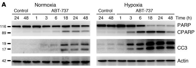
| Western blot | PARP / c-PARP / cleaved caspase 3 Hif-1a γ-H2AX / p-ATM |
|
21393866 |
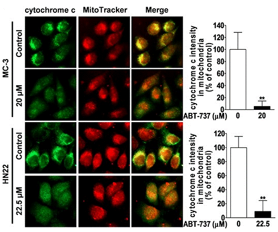
| Immunofluorescence | cytochrome C Bax Bim AIF p65 |
|
26447615 |
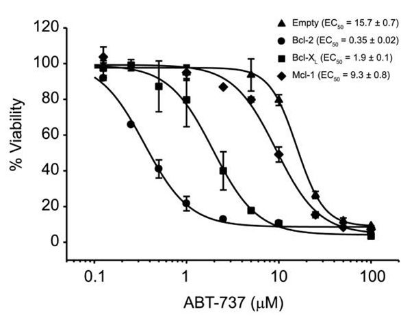
| Growth inhibition assay | Cell viability |
|
22311987 |
| ELISA | IL-6 / IL-8 |
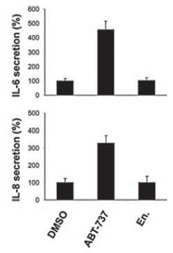
|
21084274 |
Support technique
Tel: +1-832-582-8158 Ext:3
Si vous avez dautres questions, veuillez laisser un message.
Questions fréquemment posées
Question 1:
What is the recommended method for reconstituting it for in vivo animal study?
Réponse :
For oral administration, we suggest the vehicle: 30% Propylene glycol, 5% Tween 80, 65% D5W, at up to 30mg/ml; For injection, it can be dissolved in 2% DMSO/50% PEG 300/5% Tween 80/ddH2O at 2.5 mg/ml.
Les produits sont destinés à la recherche uniquement. Non destinés à lusage humain. Nous ne vendons pas aux patients.
©Copyright 2013 Selleck Chemicals. Tous droits réservés.