alleen voor onderzoeksdoeleinden
BAY 11-7082 (BAY 11-7821) NF-κB-remmer
Cat.nr.S2913
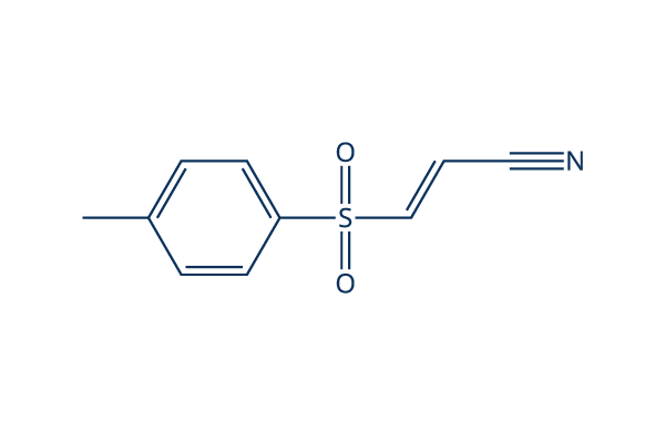
Chemische structuur
Molecuulgewicht: 207.25
Kwaliteitscontrole
| Gerelateerde doelwitten | HDAC Antioxidant ROS IκB/IKK Nrf2 AP-1 MALT NOD |
|---|---|
| Overig NF-κB Inhibitoren | DCZ0415 Omaveloxolone (RTA-408) JSH-23 QNZ (EVP4593) Caffeic Acid Phenethyl Ester SC75741 DHA (Dihydroartemisinin) Withaferin A (WFA) Andrographolide Evodiamine |
Celkweek, behandeling & werkzame concentratie
| Cellijnen | Assaytype | Concentratie | Incubatietijd | Formulering | Activiteitsbeschrijving | PMID |
|---|---|---|---|---|---|---|
| HeLa | Function Assay | 10 μM | 1.5 h | abolishes BPA induced up regulation of FN and MMP-9 | 25797437 | |
| SiHa | Function Assay | 10 μM | 1.5 h | abolishes BPA induced up regulation of FN and MMP-9 | 25797437 | |
| ARPE-19 | Function Assay | 1 μM | 0.5 h | suppresses TG-induced IL-8 promoter activation | 25593029 | |
| HCT116 | Function Assay | 5 μM | 2 h | DMSO | attenuates silymarin-induced downregulation of cyclin D1 | 25479723 |
| HMECs | Function Assay | 5 μM | 2 h | abolishes TNF-α-induced VCAM-1 expression | 25193116 | |
| A549 | Function Assay | 10 µM | 12 h | suppresses Dvl-3 induced activation of p65 | 25156800 | |
| RAW 264.7 | Function Assay | 5 μM | 1 h | inhibits TNF-α and IL-12 p40 production | 25019567 | |
| macrophages | Function Assay | 5 µM | 3 h | partially blocks YPFS-induced expression of iNOS and COX-2 | 24967898 | |
| HUVECs | Function Assay | 3-30 μM | 1 h | reduces the expression of miR-146a in a dose-dependent manner | 24863965 | |
| HeLa | Function Assay | 5 μM | 24 h | DMSO | reduces the activity of TNF-α promoter | 24657783 |
| A549 | Function Assay | 10 μM | 1 h | inhibits the increase of phospho-IκBα in PA103-infected cultures | 24612488 | |
| HUVEC | Function Assay | 20 µM | 0.5 h | DMSO | prevents the induction of EAM expression | 24551209 |
| A549RT-eto | Apoptosis Assay | 10 μM | 24 h | DMSO | accelerates FERO-mediated apoptosis | 24535083 |
| THP-1 | Function Assay | 0.1/1 μM | 0.5 h | abrogates TNF-α secretion as well as the increased secretion of IL-6 and IL-1β | 24378536 | |
| SKCXCR2 | Growth Inhibition Assay | 2 µM | 48 h | decreases cell proliferation significantly | 24376747 | |
| SKCXCR2 | Function Assay | 2 µM | 48 h | blocks the CXCL1-induced cell invasion | 24376747 | |
| OVCXCR2 | Function Assay | 2 µM | 48 h | blocks the CXCL1-induced cell invasion | 24376747 | |
| DSCs | Function Assay | 2.5 μM | 0.5 h | reverses the enhancement of CCL2/CCR2 expression of DSCs induced by IL-33 | 24344240 | |
| WPs | Function Assay | 25 μM | 5 min | suppresses ATP and vWF secretion | 24331207 | |
| A549RT-eto | Apoptosis Assay | 10 μM | 24 h | accelerates F14 extract-mediated apoptosis combined treatment with F14 | 24220725 | |
| A549RT-eto | Function Assay | 10 μM | 24 h | decreases the expression levels of NF-κB and P-gp | 24220725 | |
| FaDu | Function Assay | 2 h | inhibits p65 expression and blocks TNFα-induced TWIST expression | 24220622 | ||
| IVD | Function Assay | 10 μM | 3 d | reverses TNF-α–mediated suppression of the disc matrix macromolecules aggrecan and collagen II | 24176808 | |
| IVD | Function Assay | 10 μM | 3 d | abrogates TNF-α–induced up-regulation of ADAMTS-4 and ADAMTS-5 | 24176808 | |
| iNKT | Function Assay | 10/100 μM | 0.5 h | inhibits the induction of A2AR mRNA and other factor | 24124453 | |
| PC-3 | Function Assay | 2.5/5/10 μM | 0.5 h | blocks IGF-II-induced STS mRNA expression | 24055520 | |
| THP-1 | Function Assay | 10 μM | 1 h | abolishes the effect of rHSP27 on SR-A mRNA | 23939398 | |
| A549 | Function Assay | 1 μM | 48 h | enhances the up-regulation of IκB and subsequent decrease in Bax expression induced by combined stimulation | 23900080 | |
| A549 | Apoptosis Assay | 1 μM | 48 h | reduces the cell death induced by combined stimulation | 23900080 | |
| NCI-N87 | Growth Inhibition Assay | 10/20/30 μM | 6/24 h | suppresses cell viability significantly | 23846545 | |
| AGS | Growth Inhibition Assay | 10/20/30 μM | 6/24 h | suppresses cell viability significantly | 23846545 | |
| MGC80-3 | Growth Inhibition Assay | 10/20/30 μM | 6/24 h | suppresses cell viability significantly | 23846545 | |
| HGC-27 | Function Assay | 7.5/15/30 μM | 6 h | induces the dephosphorylation and up-regulation of IκBα | 23846545 | |
| MGC80-3 | Function Assay | 7.5/15/30 μM | 6 h | induces the dephosphorylation and up-regulation of IκBα | 23846545 | |
| HGC-27 | Apoptosis Assay | 7.5/15/30 μM | 6 h | induces apoptosis in a time- and dose-dependent manner | 23846545 | |
| HBE | Function Assay | 10μM | 3h | abolishes the increases of IL-6 expression induced by CSE | 23824089 | |
| HepG2 | Function Assay | 0.3/1/3 μM | 48 h | reduces IL6-induced PON1 expression | 23791833 | |
| THP-1 | Function Assay | 5 µM | 1 h | DMSO | inhibits MTB-induced NFκB activation | 23634218 |
| THP-1 | Growth Inhibition Assay | 5 µM | 4/8 d | DMSO | reduces the viability of intracellular MTB | 23634218 |
| MDM | Growth Inhibition Assay | 5 µM | 4/8 d | DMSO | reduces the viability of intracellular MTB | 23634218 |
| AM | Growth Inhibition Assay | 5 µM | 4/8 d | DMSO | reduces the viability of intracellular MTB | 23634218 |
| RAW 264 | Function Assay | 0.2-5 µM | 30/60/90 min | inhibits the phosphatase activity of PTP1B | 23578302 | |
| HUVEC | Function Assay | 10 μM | 0.5 h | DMSO | counteractes the loss of Tie2 mRNA | 23563632 |
| HT29 | Function Assay | 10/30/100 μM | 1 h | inhibites both TWEAK-induced p100 processing | 23527154 | |
| HT29 | Function Assay | 10/30/100 μM | 1 h | inhibits TNF-induced phosphorylation and degradation of IκBα | 23527154 | |
| MM.1S | Apoptosis Assay | 30 µM | 3 h | induces MM cell death involves necrosis | 23527154 | |
| KMS-12-BM | Apoptosis Assay | 30 µM | 3 h | induces MM cell death involves necrosis | 23527154 | |
| BAFs | Function Assay | 0.5/1 μM | 24 h | inhibits TNFα/DEX induced CYP19A1 transcripts | 23485457 | |
| SP6.5 | Function Assay | 5 μM | 2 h | decreases translocation of p65 in the nucleus | 23443086 | |
| VUP | Function Assay | 5 μM | 2 h | decreases translocation of p65 in the nucleus | 23443086 | |
| OCM1 | Function Assay | 5 μM | 2 h | decreases translocation of p65 in the nucleus | 23443086 | |
| OM431 | Function Assay | 5 μM | 2 h | decreases translocation of p65 in the nucleus | 23443086 | |
| SP6.5 | Growth Inhibition Assay | 2.5-20 μM | 24 h | IC50=5 μM, exhibits strong anti-proliferative effects in a dose-dependent manner | 23443086 | |
| VUP | Growth Inhibition Assay | 2.5-20 μM | 24 h | IC50=5 μM, exhibits strong anti-proliferative effects in a dose-dependent manner | 23443086 | |
| OCM1 | Growth Inhibition Assay | 2.5-20 μM | 24 h | IC50=5 μM, exhibits strong anti-proliferative effects in a dose-dependent manner | 23443086 | |
| OM431 | Growth Inhibition Assay | 2.5-20 μM | 24 h | IC50=5 μM, exhibits strong anti-proliferative effects in a dose-dependent manner | 23443086 | |
| SP6.5 | Apoptosis Assay | 5 μM | 24 h | induces apoptosis | 23443086 | |
| VUP | Apoptosis Assay | 5 μM | 24 h | induces apoptosis | 23443086 | |
| OCM1 | Apoptosis Assay | 5 μM | 24 h | induces apoptosis | 23443086 | |
| OM431 | Apoptosis Assay | 5 μM | 24 h | induces apoptosis | 23443086 | |
| SP6.5 | Function Assay | 5 μM | 12 h | reduces the migration | 23443086 | |
| VUP | Function Assay | 5 μM | 12 h | reduces the migration | 23443086 | |
| OCM1 | Function Assay | 5 μM | 12 h | reduces the migration | 23443086 | |
| OM431 | Function Assay | 5 μM | 12 h | reduces the migration | 23443086 | |
| HBL-1 | Growth Inhibition Assay | 3 μM | 24/48/72 h | DMSO | slows cell growth modestly | 23441730 |
| RAW 264.7 | Function Assay | 2-15 μM | 1 h | DMSO | suppresses the activation of IKK family members | 23441730 |
| IL-1R | Function Assay | 2-15 μM | 1 h | DMSO | suppresses the activation of IKK family members | 23441730 |
| RAW 264.7 | Function Assay | 15 μM | 1 h | DMSO | suppresses the activation of and JNK | 23441730 |
| IL-1R | Function Assay | 15 μM | 1 h | DMSO | suppresses the activation of and JNK | 23441730 |
| U2OS | Function Assay | 15 μM | 1 h | DMSO | prevents the LPS- or IL-1-stimulated formation of K63-pUb chains | 23441730 |
| MT‐1 | Function Assay | 8 µm | 3 h | decreases the levels of p‐STAT3 and p‐4EBP1 | 23278479 | |
| MT‐2 | Function Assay | 8 µm | 3 h | decreases the levels of p‐STAT3 and p‐4EBP1 | 23278479 | |
| MT‐1 | Function Assay | 8 µm | 3 h | decreases the levels of the p65 subunit of NF‐κB | 23278479 | |
| MT‐2 | Function Assay | 8 µm | 3 h | decreases the levels of the p65 subunit of NF‐κB | 23278479 | |
| MCF-7 | Function Assay | 2.5-15 μM | 0.5 h | DMSO | causes the gradual loss of cell adhesion | 23093227 |
| HaCaT | Function Assay | 5.0 μM | 1 h | attenuates the TCOH-induced production of IL-6 | 23041168 | |
| A549 | Function Assay | 1 h | inhibits LTA-induced SP-A mRNA production significantly | 23031213 | ||
| OA chondrocytes | Function Assay | 10 μM | 1 h | blocks the AGE-BSA-induced gene/protein expression of GRP78 or COX-2 (p<0.05) | 22982228 | |
| RAW264.7 | Function Assay | 15 μM | 15-120 min | blocks the production of NO, PGE2, and TNF-α | 22745523 | |
| RAW264.7 | Growth Inhibition Assay | 5-30 μM | 24 h | inhibits cell growth in a dose-dependent manner | 22745523 | |
| HBL6 | Apoptosis Assay | 0.5/5/25 μM | 6/24 h | decreases cell viability and leeads to apoptosis in a dose-dependent manner | 22074820 | |
| HT29 | Function Assay | 1-10 μM | 10 h | increases HO-1 mRNA and protein expression | 21620964 | |
| Ca9–22 | Apoptosis Assay | 10 μM | 1 h | completely inhibits ALA-PDT-induced apoptosis | 21138480 | |
| Ca9–22 | Function Assay | 10 μM | 1 h | completely abrogates the ALA-PDT-induced JNK activation | 21138480 | |
| A-549 | Growth Inhibition Assay | 10 μM | 24/48 h | inhibits cell growth in a time-dependent manner | 20866043 | |
| AP | Function Assay | 5/10 μM | 48 h | downregulates the BAD protein level a dose-dependent manner | 20596645 | |
| AQ1 | Function Assay | 5/10 μM | 48 h | downregulates the BAD protein level a dose-dependent manner | 20596645 | |
| AP | Function Assay | 20 μM | 4/8 h | downregulates the BAD protein level a time-dependent manner | 20596645 | |
| AQ1 | Function Assay | 20 μM | 4/8 h | downregulates the BAD protein level a time-dependent manner | 20596645 | |
| THP-1 | Function Assay | 5 μM | 0.5 h | attenuates the LPS-induced p-IκBα protein by 72% | 20309718 | |
| K562 | Growth Inhibition Assay | 2-30 μM | 24 h | IC50=8 μM,inhibits cell growth in a dose-dependent manner | 19646807 | |
| Jurket | Growth Inhibition Assay | 2-30 μM | 24 h | IC50=7.1 μM, inhibits cell growth in a dose-dependent manner | 19646807 | |
| U937 | Growth Inhibition Assay | 2-30 μM | 24 h | IC50=10.5 μM, inhibits cell growth in a dose-dependent manner | 19646807 | |
| PBMC | Growth Inhibition Assay | 2-30 μM | 24 h | IC50=40.2 μM, inhibits cell growth in a dose-dependent manner | 19646807 | |
| K562 | Apoptosis Assay | 2-20 μM | 24 h | induces a dose-dependent apoptosis | 19646807 | |
| THP1 | Cytotoxicity assay | 72 hrs | Cytotoxicity against human THP1 cells assessed as reduction in cell viability after 72 hrs by MTT assay, TC50 = 1.5 μM. | 28410442 | ||
| RAW264.7 | Function assay | 6 hrs | Inhibition of LPS-induced NF-kappaB activation in mouse RAW264.7 cells treated 30 mins before LPS challenge measured after 6 hrs by luciferase reporter gene assay, IC50 = 1.72 μM. | 24315191 | ||
| HEK293 | Function assay | 6 hrs | Inhibition of TNF-alpha-induced NF-kappaB activity in HEK293 cells after 6 hrs by luciferase reporter gene assay, IC50 = 2 μM. | 24533857 | ||
| HEK293 | Function assay | 6 hrs | Inhibition of TNF-alpha-induced NF-kappaB activity in HEK293 cells after 6 hrs by luciferase reporter gene assay, IC50 = 2 μM. | 24992702 | ||
| HEK293 | Function assay | 6 hrs | Inhibition of TNFalpha-induced NF-kappaB activity (unknown origin) transfected in HEK293 cells after 6 hrs by luciferase reporter gene assay, IC50 = 2 μM. | 26343828 | ||
| HEK293 | Function assay | 6 hrs | Inhibition of TNFalpha-induced NF-kappaB activity expressed in human HEK 293 cells after 6 hrs by luciferase reporter gene assay, IC50 = 2 μM. | 22850207 | ||
| HEK293 | Function assay | 6 hrs | Inhibition of TNFalpha-induced human NFkappaB activity in HEK293 cells incubated for 6 hrs followed by compound wash out measured after 5 mins by by luciferase assay, IC50 = 2.01 μM. | 22712432 | ||
| HEK293 | Cytotoxicity assay | Cytotoxicity against HEK293 cells, IC50 = 3.8 μM. | 24533857 | |||
| HEK293 | Function assay | 6 hrs | Inhibition of TNFalpha-induced NFkappaB (unknown origin) activation expressed in HEK293 cells after 6 hrs by luciferase reporter gene assay, IC50 = 5 μM. | 23316950 | ||
| HEK293 | Function assay | Effect on Cdc2 expressed in HEK293 cells assessed as effect on Cdc2:Cdc25C interaction complexes in presence of camptothecin by EYFP and/or YFP Venus fragment based reporter gene assay | 16680159 | |||
| HEK293 | Function assay | 20 uM | 24 hrs | Inhibition of TNF-alpha stimulated NFkappaB transactivation in HEK293 cells at 20 uM measured after 24 hrs by dual luciferase reporter gene assay | 27736063 | |
| RAW264.7 | Function assay | 20 uM | 1 hr | Inhibition of LPS-induced NFkB activation in mouse RAW264.7 cells assessed as reduction in nuclear translocation of p65 at 20 uM preincubated for 1 hr followed by LPS stimulation measured after 3 hrs by Western blot method | 28667873 | |
| RAW264.7 | Function assay | 20 uM | 6 hrs | Inhibition of LPS-induced NF-kappaB activation in mouse RAW264.7 cells at 20 uM treated 30 mins before LPS challenge measured after 6 hrs by luciferase reporter gene assay | 24315191 | |
| RAW264.7 | Antinflammatory assay | 20 uM | 18 hrs | Antinflammatory activity in mouse RAW264.7 cells assessed as inhibition of LPS-induced nitric oxide production at 20 uM treated 30 mins before LPS challenge measured after 18 hrs by Griess assay | 24315191 | |
| THP1 | Antinflammatory assay | 5 uM | 24 hrs | Antiinflammatory activity in human THP1 cells assessed as inhibition of TPA/ionomycin-induced extracellular IL-1beta level at 5 uM incubated 1 hr prior to TPA/ionomycin challenge measured after 24 hrs by ELISA | 24400858 | |
| THP1 | Antinflammatory assay | 5 uM | 24 hrs | Antiinflammatory activity in human THP1 cells assessed as inhibition of TPA/ionomycin-induced extracellular TNF-alpha production at 5 uM incubated 1 hr prior to TPA/ionomycin challenge measured after 24 hrs by ELISA | 24400858 | |
| THP1 | Antinflammatory assay | 5 uM | 24 hrs | Antiinflammatory activity in human THP1 cells assessed as inhibition of TPA/ionomycin-induced intracellular proIL-1beta level at 5 uM incubated 1 hr prior to TPA/ionomycin challenge measured after 24 hrs by ELISA | 24400858 | |
| THP1 | Antinflammatory assay | 5 uM | 24 hrs | Antiinflammatory activity in human THP1 cells assessed as inhibition of TPA/ionomycin-induced intracellular IL-1beta level at 5 uM incubated 1 hr prior to TPA/ionomycin challenge measured after 24 hrs by ELISA | 24400858 | |
| RAW264.7 | Function assay | 0.3 ug/ml | 12 hrs | Inhibition of LPS-induced NF-kB p65 phosphorylation in mouse RAW264.7 cells at 0.3 ug/ml preincubated for 12 hrs followed by LPS stimulation for 3 hrs by Western blot method | 28284806 | |
| RAW264.7 | Function assay | 0.3 ug/ml | 12 hrs | Inhibition of LPS-induced NF-kB p65 activation in mouse RAW264.7 cells at 0.3 ug/ml preincubated for 12 hrs followed by LPS stimulation for 3 hrs by DAPI staining based inverted fluorescence microscopic method | 28284806 | |
| RAW264.7 | Function assay | 0.3 ug/ml | 12 hrs | Inhibition of NF-kB p65 in mouse RAW264.7 cells assessed as reduction in LPS-induced iNOS expression at 0.3 ug/ml preincubated for 12 hrs followed by LPS stimulation for 3 hrs by Western blot method | 28284806 | |
| RAW264.7 | Function assay | 0.3 ug/ml | 12 hrs | Inhibition of NF-kB p65 in mouse RAW264.7 cells assessed as reduction in LPS-induced COX2 expression at 0.3 ug/ml preincubated for 12 hrs followed by LPS stimulation for 3 hrs by Western blot method | 28284806 | |
| HEK293 | Function assay | 20 uM | 24 hrs | Inhibition of TNFalpha-induced NFkappaB activation in HEK293 cells at 20 uM after 24 hrs by dual luciferase reporter gene assay | 28873303 | |
| RAW264.7 | Function assay | 20 uM | 2 hrs | Inhibition of NFkappaB nuclear translocation in LPS-stimulated mouse RAW264.7 cells at 20 uM pretreated for 2 hrs followed by LPS-induction by DAPI-staining based immunofluorescence microscopic method | 29759725 | |
| BGC823 | Function assay | 5 uM | 12 hrs | Inhibition of colony formation in human BGC823 cells at 5 uM treated for 12 hrs followed by incubation in drug free medium for 14 days by crystal violet staining based assay | 28881286 | |
| SGC7901 | Function assay | 5 uM | 12 hrs | Inhibition of colony formation in human SGC7901 cells at 5 uM treated for 12 hrs followed by incubation in drug free medium for 14 days by crystal violet staining based assay | 28881286 | |
| RAW264.7 | Function assay | 10 uM | 2 hrs | Inhibition of LPS-induced IL-6 mRNA expression in mouse RAW264.7 cells at 10 uM pre-incubated for 2 hrs before LPS stimulation for 24 hrs by qRT-PCR method | 27038497 | |
| RAW264.7 | Function assay | 10 uM | 2 hrs | Inhibition of LPS-induced IL-1beta mRNA expression in mouse RAW264.7 cells at 10 uM pre-incubated for 2 hrs before LPS stimulation for 24 hrs by qRT-PCR method | 27038497 | |
| RAW264.7 | Function assay | 10 uM | 2 hrs | Inhibition of LPS-induced iNOS mRNA expression in mouse RAW264.7 cells at 10 uM pre-incubated for 2 hrs before LPS stimulation for 24 hrs by qRT-PCR method | 27038497 | |
| Klik om meer experimentele gegevens over cellijnen te bekijken | ||||||
Chemische informatie, opslag en stabiliteit
| Molecuulgewicht | 207.25 | Formule | C10H9NO2S |
Opslag (vanaf de datum van ontvangst) | |
|---|---|---|---|---|---|
| CAS-nr. | 19542-67-7 | SDF downloaden | Opslag van stamoplossingen |
|
|
| Synoniemen | BAY 11-7821 | Smiles | CC1=CC=C(C=C1)S(=O)(=O)C=CC#N | ||
Oplosbaarheid
|
In vitro |
DMSO
: 41 mg/mL
(197.82 mM)
Ethanol : 10 mg/mL Water : Insoluble |
Molariteitscalculator
|
In vivo |
|||||
In vivo formulatiecalculator (heldere oplossing)
Stap 1: Voer onderstaande informatie in (Aanbevolen: een extra dier om rekening te houden met verlies tijdens het experiment)
Stap 2: Voer de in vivo formulering in (Dit is alleen de calculator, geen formulering. Neem eerst contact met ons op als er geen in vivo formulering is in de sectie oplosbaarheid.)
Berekeningsresultaten:
Werkconcentratie: mg/ml;
Methode voor het bereiden van DMSO-moedervloeistof: mg geneesmiddel vooropgelost in μL DMSO ( Concentratie moedervloeistof mg/mL, Neem eerst contact met ons op als de concentratie de DMSO-oplosbaarheid van de batch van het geneesmiddel overschrijdt. )
Methode voor het bereiden van in vivo formulering: Neem μL DMSO moedervloeistof, voeg daarna toeμL PEG300, mengen en verhelderen, daarna toevoegenμL Tween 80, mengen en verhelderen, daarna toevoegen μL ddH2O, mengen en verhelderen.
Methode voor het bereiden van in vivo formulering: Neem μL DMSO moedervloeistof, voeg daarna toe μL Maïsolie, mengen en verhelderen.
Opmerking: 1. Zorg ervoor dat de vloeistof helder is voordat u het volgende oplosmiddel toevoegt.
2. Zorg ervoor dat u het/de oplosmiddel(en) in de juiste volgorde toevoegt. U moet ervoor zorgen dat de verkregen oplossing, bij de vorige toevoeging, een heldere oplossing is voordat u verdergaat met het toevoegen van het volgende oplosmiddel. Fysieke methoden zoals vortexen, ultrasoon of een warmwaterbad kunnen worden gebruikt om het oplossen te bevorderen.
Werkingsmechanisme
| Targets/IC50/Ki |
E2-conjugating enzymes
(Cell-free assay) USP7
(Cell-free assay) 0.19 μM
USP21
(Cell-free assay) 0.96 μM
USP6
(Cell-free assay) 1.7 μM
IκBα phosphorylation
(Tumor cells) 10 μM
|
|---|---|
| In vitro |
BAY 11-7082 heft de NF-κB DNA-binding volledig en specifiek op, waardoor de NF-κB-induceerbare cytokine IL-6 wordt gedownreguleerd en apoptose wordt geïnduceerd. Deze verbinding (< 8 μM) is in staat om zowel de basale als de TNFα gestimuleerde NFκB luciferase-activiteit effectief te remmen op een dosisafhankelijke manier. Het (8 μM) remt sterk de proliferatiesnelheid in NCI-H1703 cellen. Deze verbinding (5 μM) vermindert snel en efficiënt de DNA-binding van NF-kappaB in HTLV-I-geïnfecteerde T-cellijnen en down-reguleert de expressie van het anti-apoptotische gen, Bcl-x(L), terwijl het weinig effect heeft op de DNA-binding van een andere transcriptiefactor, AP-1. Deze chemisch geïnduceerde apoptose van primaire ATL-cellen is prominenter dan die van normale perifere bloedmononucleaire cellen, en apoptose van deze cellen is ook geassocieerd met down-regulatie van NF-kappaB-activiteit. Het (5 μM) induceert selectief apoptose van HTLV-I-geïnfecteerde T-cellijnen geassocieerd met down-regulatie van de expressie van cycline D1, cycline D2 en Bcl-xL. Deze verbinding (100 μM) voorkomt de nucleaire translocatie van p65, uitgelokt door NMDA, en de NMDA-geïnduceerde toename van NF-κB-binding in muizen hippocampuscoupes. Het voorkomt NMDA-toxiciteit in het CA1-gebied van hippocampuscoupes met 40% neuroprotectie bij 20 μM en 70% neuroprotectie bij 100 μM. Deze chemische stof remt bij alle geteste concentraties significant de NF-κB p65 DNA-bindende activiteit in vetweefsel, terwijl het in skeletspieren, bij 50 μM en 100 μM, significant de NF-κB p65 DNA-bindende activiteit remt. Het (100 μM) vermindert IKK-β-eiwit in menselijk vetweefsel en skeletspieren. Deze verbinding (100 μM) vermindert significant de afgifte van TNF-α uit vetweefsel, terwijl de afgifte van IL-6 en IL-8 significant wordt geremd bij alle geteste concentraties van deze chemische stof. Het (50 μM) vermindert significant de afgifte van TNF-α, IL-6 en IL-8 in skeletspieren. Deze verbinding blijkt ook de E2-conjugerende enzymen Ubc (ubiquitine conjugerende) 13 en UbcH7 en de E3-ligase LUBAC (lineair ubiquitine-assemblagecomplex) te inactiveren, en induceert zo de dood van B-cellymfoom- en leukemische T-cellen. |
| In vivo |
BAY 11-7082, een NF-κB-remmer, induceert apoptose en S-fase-arrest in maagkankercellen. |
Referenties |
|
Toepassingen
| Methoden | Biomarkers | Afbeeldingen | PMID |
|---|---|---|---|
| Western blot | NF-κB p-IKKβ/ IκBα p-IRAK4 / IRAK4 NF-κB p-IKKβ/ IκBα p-IRAK4 / IRAK4 |
|
31332209 |
| Growth inhibition assay | Cell viability Cell viability |
|
31332209 |
Technische ondersteuning
Tel: +1-832-582-8158 Ext:3
Als u nog andere vragen heeft, laat dan een bericht achter.
Producten zijn uitsluitend voor onderzoeksdoeleinden. Niet voor menselijk gebruik. Wij verkopen niet aan patiënten.
©Copyright 2013 Selleck Chemicals. Alle rechten voorbehouden.