alleen voor onderzoeksgebruik
ApoA1 Antibody [L13J2]
Cat.nr.: F5020
Toepassing:
Reactiviteit:
-
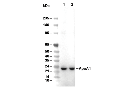 Lane 1: Human small intestine, Lane 2: Human plasma
Experimentbenodigdheden
WB
Recommended wet transfer conditions: 200 mA, 60 min.
Recommended wet transfer conditions: 200 mA, 60 min.
Gebruiksinformatie
| Verdunning |
|---|
|
| Toepassing |
|---|
| WB |
| Reactiviteit |
|---|
| Human |
| Bron |
|---|
| Mouse Monoclonal Antibody |
| Opslagbuffer |
|---|
| PBS, pH 7.2+50% Glycerol+0.05% BSA+0.01% NaN3 |
| Opslag (vanaf ontvangstdatum) |
|---|
| -20°C (avoid freeze-thaw cycles), 2 years |
| Voorspeld MW |
|---|
| 25 kDa |
| Positieve controle | HepG2 concentrated media |
|---|---|
| Negatieve controle | HepG2 cells |
Experimentele methoden
| WB |
|---|
Experimental Protocol:
Sample preparation
1. Tissue: Lyse the tissue sample by adding an appropriate volume of ice-cold RIPA/NP-40 Lysis Buffer (containing Protease Inhibitor Cocktail),and homogenize the tissue at a low temperature. 2. Adherent cell: Aspirate the culture medium and wash the cells with ice-cold PBS twice. Lyse the cells by adding an appropriate volume of RIPA/NP-40 Lysis Buffer (containing Protease Inhibitor Cocktail) and put the sample on ice for 5 min. 3. Suspension cell: Transfer the culture medium to a pre-cooled centrifuge tube. Centrifuge and aspirate the supernatant. Wash the cells with ice-cold PBS twice. Lyse the cells by adding an appropriate volume of RIPA/NP-40 Lysis Buffer (containing Protease Inhibitor Cocktail) and put the sample on ice for 5 min. 4. Place the lysate into a pre-cooled microcentrifuge tube. Centrifuge at 4°C for 15 min. Collect the supernatant;
5. Remove a small volume of lysate to determine the protein concentration;
6. Combine the lysate with protein loading buffer. Boil 20 µL sample under 95-100°C for 5 min. Centrifuge for 5 min after cool down on ice.
Electrophoretic separation
1. According to the concentration of extracted protein, load appropriate amount of protein sample and marker onto SDS-PAGE gels for electrophoresis. Recommended separating gel (lower gel) concentration: 10%. Reference Table for Selecting SDS-PAGE Separation Gel Concentrations 2. Power up 80V for 30 minutes. Then the power supply is adjusted (110 V~150 V), the Marker is observed, and the electrophoresis can be stopped when the indicator band of the predyed protein Marker where the protein is located is properly separated. (Note that the current should not be too large when electrophoresis, too large current (more than 150 mA) will cause the temperature to rise, affecting the result of running glue. If high currents cannot be avoided, an ice bath can be used to cool the bath.)
Transfer membrane
1. Take out the converter, soak the clip and consumables in the pre-cooled converter;
2. Activate PVDF membrane with methanol for 1 min and rinse with transfer buffer;
3. Install it in the order of "black edge of clip - sponge - filter paper - filter paper - glue -PVDF membrane - filter paper - filter paper - sponge - white edge of clip"; 4. The protein was electrotransferred to PVDF membrane. ( 0.45 µm PVDF membrane is recommended ) Reference Table for Selecting PVDF Membrane Pore Size Specifications Recommended conditions for wet transfer: 200 mA, 60 min. ( Note that the transfer conditions can be adjusted according to the protein size. For high-molecular-weight proteins, a higher current and longer transfer time are recommended. However, ensure that the transfer tank remains at a low temperature to prevent gel melting.)
Block
1. After electrotransfer, wash the film with TBST at room temperature for 5 minutes;
2. Incubate the film in the blocking solution for 1 hour at room temperature;
3. Wash the film with TBST for 3 times, 5 minutes each time.
Antibody incubation
1. Use 5% skim milk powder to prepare the primary antibody working liquid (recommended dilution ratio for primary antibody 1:1000), gently shake and incubate with the film at 4°C overnight; 2. Wash the film with TBST 3 times, 5 minutes each time;
3. Add the secondary antibody to the blocking solution and incubate with the film gently at room temperature for 1 hour;
4. After incubation, wash the film with TBST 3 times for 5 minutes each time.
Antibody staining
1. Add the prepared ECL luminescent substrate (or select other color developing substrate according to the second antibody) and mix evenly;
2. Incubate with the film for 1 minute, remove excess substrate (keep the film moist), wrap with plastic film, and expose in the imaging system. |
Biologische beschrijving
| Specificiteit |
|---|
| ApoA1 Antibody [L13J2] detects endogenous levels of total ApoA1 protein. |
| Subcellulaire lokalisatie |
|---|
| Amyloid, HDL, Secreted |
| Uniprot ID |
|---|
| P02647 |
| Kloon |
|---|
| L13J2 |
| Synoniem |
|---|
| Apolipoprotein A-I; Apo-AI; ApoA-I; Apolipoprotein A1; APOA1 |
| Achtergrond |
|---|
| Apolipoprotein A-I (ApoA-I) is the primary structural and functional protein of high-density lipoprotein (HDL), synthesized mainly in the liver and intestine to mediate reverse cholesterol transport by facilitating ABCA1-dependent cholesterol efflux from peripheral tissues to the liver for excretion, thereby offering protection against atherosclerosis and cardiovascular disease. The mature 243-amino-acid ApoA-I polypeptide comprises 10 tandem repeats of amphipathic α-helices, with proline residues providing flexibility for lipid binding; its N-terminal region stabilizes the lipid-free form, the central domain (140–170) activates lecithin-cholesterol acyltransferase (LCAT) for cholesterol esterification and HDL maturation, and the C-terminal domain (181–243) enables high-affinity phospholipid binding for nascent HDL formation. ApoA-I serves as a cofactor for LCAT, interacts with ABCA1 and SR-BI receptors to promote lipid efflux, and exhibits strong anti-inflammatory and antioxidant actions by neutralizing lipopolysaccharides and inhibiting vascular adhesion molecule expression. Low ApoA-I levels are an independent risk predictor for coronary artery disease, while APOA1 mutations cause familial HDL deficiency syndromes or hereditary amyloidosis, leading to accelerated atherosclerosis or organ dysfunction from protein deposits, with additional links to Alzheimer’s disease and diabetes through impaired lipid homeostasis and chronic inflammation. |
| Referenties |
|---|
|
Technische ondersteuning
Tel: +1-832-582-8158 Ext:3
Als u nog andere vragen heeft, laat dan een bericht achter.
Producten zijn uitsluitend voor onderzoeksdoeleinden. Niet voor menselijk gebruik. Wij verkopen niet aan patiënten.
©Copyright 2013 Selleck Chemicals. Alle rechten voorbehouden.