Vadimezan (DMXAA)

CatalogusnummerS1537 Batch:S153705

Afdrukken

Technische gegevens

Formule

C17H14O4

Moleculair gewicht 282.29 CAS-nr. 117570-53-3
Oplosbaarheid (25°C)* In vitro DMSO 4 mg/mL (14.16 mM)
Water Insoluble
Ethanol Insoluble
In vivo (Voeg oplosmiddelen afzonderlijk en in volgorde toe aan het product.)
Homogeneous suspension
CMC-NA
≥5mg/ml Taking the 1 mL working solution as an example, add 5 mg of this product to 1 ml of CMC-Na solution, mix evenly to obtain a homogeneous suspension with a final concentration of 5 mg/ml.
* <1 mg/ml betekent licht oplosbaar of onoplosbaar.
* Houd er rekening mee dat Selleck de oplosbaarheid van alle verbindingen intern test en de werkelijke oplosbaarheid enigszins kan afwijken van gepubliceerde waarden. Dit is normaal en is te wijten aan lichte batch-tot-batch variaties.
* Verzending op kamertemperatuur (Stabiliteitstests tonen aan dat dit product zonder koelmaatregelen kan worden verzonden.)

Voorbereiden van stamoplossingen

Biologische activiteit

Beschrijving Vadimezan (DMXAA) is een vasculair verstorend agens (VDA) en een competitieve remmer van DT-diaphorase met een Ki van 20 μM en een IC50 van 62,5 μM in celvrije assays, respectievelijk. Het is tevens een STING agonist met potentiële antineoplastische activiteit, die in vitro krachtig IFN-β maar relatief lage TNF-α expressie induceert. Deze verbinding heeft Antiviral activiteit. Fase 3.
Doelen
DT-diaphorase
(Cell-free assay)
DT-diaphorase
(Cell-free assay)
20 μM(Ki) 20 μM(Ki)
In vitro In DLD-1 menselijke darmcarcinoomcellen remt Vadimezan (DMXAA) de DT-diaphorase-activiteit zonder significante effecten op de activiteit van cytochroom b5-reductase en cytochroom P450-reductase. De combinatie van menadion en deze verbinding leidt tot een toename van de antiproliferatieve activiteit van DLD-1-cellen. Als Antiviral middel remt het VSV-geïnduceerde cytotoxiciteit en influenzavirusreplicatie in RAW 264.7-macrofagen. Een recente studie toont aan dat DMXAA niet-immuungemedieerde remmende effecten heeft tegen verschillende kinaseleden van VEGFR (vascular endothelial growth factor receptor), zoals VEGFR2-signalering in menselijke navelstrengendotheelcellen.
In vivo Vadimezan (DMXAA)-behandeling beschermt C57BL/6J-muizen significant die i.n. zijn geïnfecteerd met 200 p.f.u. muis-aangepast H1N1-influenzavirus PR8 met 60% overleving, terwijl de controlegroep slechts 20% overleving vertoonde. Deze verbinding vertraagt de tumorgroei die wordt geïnduceerd door chemische carcinogenen significant, verlengt de tijd tot tumordubbeling en verlengt de tijd vanaf behandeling tot euthanasie. Na behandeling werden de mediane tumordubbelingstijd, de mediane tumortriplingstijd en de mediane tijd vanaf behandeling tot euthanasie bij tumor-dragende dieren met respectievelijk ongeveer 4,4-, 1,8- en 2,7-voudig verhoogd.

Protocol (uit referentie)

Kinase-assay:[1]
  • DT-diaforase activiteit en kinetische analyse van enzymremming

    Gezuiverde DT-diaforase enzymactiviteit wordt gemeten door de reductie van cytochroom c te meten bij 550 nm op een Beckman DU 650 spectrofotometer. Elke assay bevat cytochroom c (70 μM), NADH (variabele concentraties), gezuiverde DT-diaforase (0,032 μg) en menadion (variabele concentraties) in een eindvolume van 1 ml Tris-HCl buffer (50 mM, pH 7,4) met 0,14% BSA. De reactie wordt gestart door de toevoeging van NADH. De reductiesnelheden worden berekend over het initiële deel van de reactiecurve (30 seconden), en de resultaten worden uitgedrukt in μmol cytochroom c gereduceerd/min/mg eiwit met behulp van een molaire extinctiecoëfficiënt van 21,1 mM−1 cm−1 voor gereduceerd cytochroom c. Enzymassays worden uitgevoerd bij kamertemperatuur en alle reacties worden in drievoud uitgevoerd. Remming van de gezuiverde DT-diaforase activiteit wordt uitgevoerd door de opname van Vadimezan (DMXAA) in verschillende concentraties in de reactie, en remmingskenmerken worden bepaald door de concentratie van NADH (constante menadion) of menadion (constante NADH) bij verschillende concentraties van deze verbinding te variëren. Ki-waarden worden verkregen door 1/V uit te zetten tegen. De activiteit van DT-diaforase in DLD-1 cellen wordt bepaald door de dicumarol-gevoelige reductie van DCPIP te meten bij 600 nm. Kortom, DLD-1 cellen in de midden-exponentiële groeifase worden geoogst door afschrapen in ijskoude buffer (Tris-HCl, 25 mM, pH 7,4 en 250 mM sucrose) en gesoniceerd op ijs. Enzymassaycondities zijn 2 mM NADH, 40 μM DCPIP, 20 μL dicumarol (indien nodig) in een eindvolume van 1 ml Tris-HCl (25 mM, pH 7,4) met BSA (0,7 mg/ml). Resultaten worden uitgedrukt als de dicumarol-gevoelige reductie van DCPIP met behulp van een molaire extinctiecoëfficiënt van 21 mM−1 cm−1. Eiwitniveaus worden bepaald met behulp van de Bradford-assay.

Celassay:[1]
  • Cellijnen

    DLD-1 and H460 cells

  • Concentraties

    0-2 mM

  • Incubatietijd

    96 hours

  • Methode

    DLD-1 human colon carcinoma and H460 human non-small cell lung carcinoma cells are routinely maintained as monolayer cultures in RPMI 1640 culture medium supplemented with foetal calf serum (10%), sodium pyruvate (2 mM), penicillin/streptomycin (50 IU mL−1/50 μg mL-1) and l-glutamine (2 mM). Chemosensitivity is assessed using the MTT assay and all assays are performed under aerobic conditions. Briefly, cells are plated into each well of a 96-well culture plate and incubated overnight in an atmosphere containing 5% CO2. Culture medium is completely removed from each well and replaced with medium containing a range of drug concentrations. In the case of menadione alone, the duration of drug exposure is 1 hour, after which the cells are washed twice with Hanks' balanced salt solution prior to the addition of 200 μL fresh RPMI 1640 medium to each well of the plate. For Vadimezan (DMXAA) alone, the duration of exposure is 3 hours. Following a four-day incubation, cell survival is determined using the MTT assay. For combinations of this compound with menadione, cells are initially set up and a non-toxic (>95% cell survival) concentration of it is selected. Cells are then initially exposed to DMXAA (2 mM) for a period of 2 hours, following which the medium is removed and replaced with medium containing the inhibitor (at a constant concentration of 2 mM) and menadione (at a range of drug concentrations). Following a further 7-hour incubation, cells are washed twice with Hanks' balanced salt solution prior to the addition of growth medium.

Dierstudie:[4]
  • Dierlijke modellen

    Chemical carcinogen (NMU) is injected into female Wistar rats.

  • Doseringen

    ≤300 mg/kg

  • Toediening

    Administered via i.p.

Referenties

  • https://pubmed.ncbi.nlm.nih.gov/10423172/
  • https://pubmed.ncbi.nlm.nih.gov/21084628/
  • https://pubmed.ncbi.nlm.nih.gov/22142330/
  • https://pubmed.ncbi.nlm.nih.gov/16944150/

Klantproductvalidatie

(B and C) sh-scrambled or sh-ck2a–transducted L929 cells (B) and Raw cells (C) were stimulated by DMXAA (100 μg/ml) for various times. Cytosolic and nuclear extracts were prepared as described in Materials and Methods. Five percent of the cytosolic proteins and 20% of the nuclear proteins were resolved by 10% SDS-PAGE. Subsequently, immunoblotting was conducted by indicated Abs. The amounts of Tubulin and Lamin B1 in cytosol versus nuclei detected by respective Abs were used as internal control for fractionation.

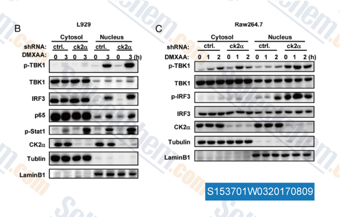
Gegevens van [ , , J Immunol, 2015, 194:4477-4488 ]

(E) WT and ASC−/− macrophages were stimulated with 1 μg/ml of tumor-derived DNA in the presence of lipofectamine or 50 μg/ml of DMXAA at different time points. Whole cell extracts were analysed with antibodies against pTBK1, total TBK1, pIRF3, total IRF3 and GAPDH.

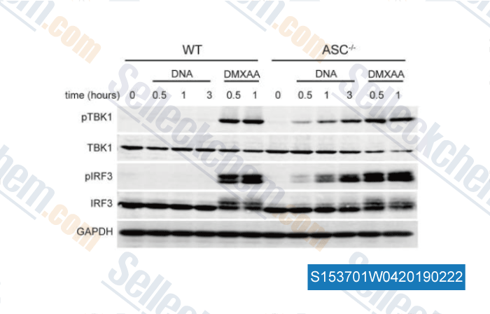
Gegevens van [ , , J Immunol, 2016, 196(7):3191-8 ]

(B and C) sh-scrambled or sh-ck2α–transducted L929 cells (B) and Raw cells (C) were stimulated by DMXAA (100 μg/ml) for various times. Cytosolic and nuclear extracts were prepared as described in Materials and Methods. Five percent of the cytosolic proteins and 20% of the nuclear proteins were resolved by 10% SDS-PAGE. Subsequently, immunoblotting was conducted by indicated Abs. The amounts of Tubulin and Lamin B1 in cytosol versus nuclei detected by respective Abs were used as internal control for fractionation.

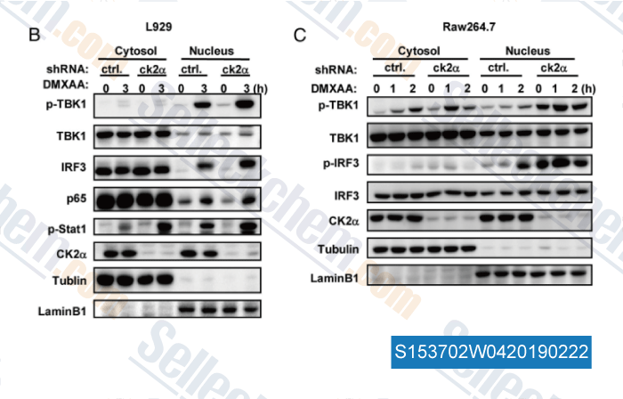
Gegevens van [ , , J Immunol, 2015, 194(9):4477-88 ]

(b-e) HEK293T cells were transiently transfected with empty vector (b), plasmids encoding hSTING(H232) (c), hSTING(R232) (d), or mSTING (e) together with IFNβ-luciferase reporter. After 24 h, cells were transfected with 2′,3′‐cGAMP (2 μg/mL) or stimulated with CMA (50 μg/mL), DMXAA (50 μM) and α-mangostin. Luciferase activity was measured 24 h after stimulation. Error bars represent the SD of independent experiments (n=3); *P<0.05, **P<0.01, ***P<0.001 (Student

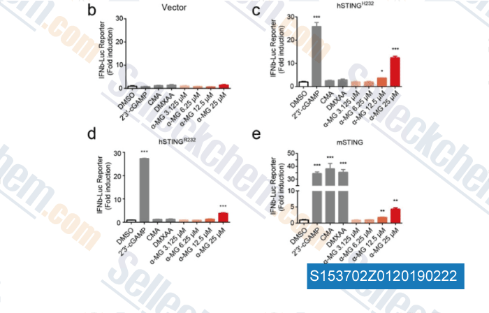
Gegevens van [ , , ChemMedChem, 2018, 13(19):2057-2064 ]

Sellecks Vadimezan (DMXAA) Is geciteerd door 46 Publicaties

An oral tricyclic STING agonist suppresses tumor growth through remodeling of the immune microenvironment [ Cell Chem Biol, 2025, 32(2):280-290.e14] PubMed: 39904339
LAPTM5 exacerbates STING-mediated inflammation induced by LL-37 through stabilizing STING in rosacea [ Commun Biol, 2025, 8(1):1470] PubMed: 41087666
Profile of STING agonist and inhibitor research: a bibliometric analysis [ Front Pharmacol, 2025, 16:1528459] PubMed: 40008133
C9orf72 Alleviates DSS‑Induced Ulcerative Colitis via the cGAS-STING Pathway [ Immun Inflamm Dis, 2025, 13(1):e70139] PubMed: 39873292
YTHDF2 in peritumoral hepatocytes mediates chemotherapy-induced antitumor immune responses through CX3CL1-mediated CD8+ T cell recruitment [ Mol Cancer, 2024, 23(1):186] PubMed: 39237909
S-nitrosothiol homeostasis maintained by ADH5 facilitates STING-dependent host defense against pathogens [ Nat Commun, 2024, 15(1):1750] PubMed: 38409248
Mitochondrial DNA-boosted dendritic cell-based nanovaccination triggers antitumor immunity in lung and pancreatic cancers [ Cell Rep Med, 2024, 5(7):101648] PubMed: 38986624
FMT rescues mice from DSS-induced colitis in a STING-dependent manner [ Gut Microbes, 2024, 16(1):2397879] PubMed: 39324491
TBK1-Zyxin signaling controls tumor-associated macrophage recruitment to mitigate antitumor immunity [ EMBO J, 2024, 10.1038/s44318-024-00244-9] PubMed: 39304793
ISGylation by HERCs facilitates STING activation [ Cell Rep, 2024, 43(5):114135] PubMed: 38652662

RETOURBELEID
Selleck Chemicals onvoorwaardelijke retourbeleid zorgt voor een soepele online winkelervaring voor onze klanten. Als u op enigerlei wijze ontevreden bent met uw aankoop, kunt u elk artikel(en) binnen 7 dagen na ontvangst retourneren. In geval van problemen met de productkwaliteit, zowel protocolgerelateerde als productgerelateerde problemen, kunt u elk artikel(en) binnen 365 dagen na de oorspronkelijke aankoopdatum retourneren. Volg de onderstaande instructies bij het retourneren van producten.

VERZENDING EN OPSLAG
Selleck producten worden bij kamertemperatuur vervoerd. Als u het product op kamertemperatuur ontvangt, wees dan gerust, de Selleck kwaliteitsinspectieafdeling heeft experimenten uitgevoerd om te controleren of de normale temperatuurplaatsing van één maand de biologische activiteit van poederproducten niet beïnvloedt. Na ontvangst dient u het product op te slaan volgens de vereisten beschreven in het gegevensblad. De meeste Selleck producten zijn stabiel onder de aanbevolen omstandigheden.

NIET VOOR HUMANE, VETERINAIRE DIAGNOSTISCHE OF THERAPEUTISCHE DOELEINDEN.