|
Hoe te citeren 1. Voor in-tekst citatie (materialen en methoden): 2. Voor de tabel met belangrijke bronnen: |
||
|
Gratis nummer: (877) 796-6397 -- Alleen VS en Canada -- |
Fax: +1-832-582-8590 Bestellingen: +1-832-582-8158 |
Technische ondersteuning: +1-832-582-8158 Ext:3 Vermeld uw bestelnummer in de e-mail. Wij streven ernaar alle e-mailvragen binnen één werkdag te beantwoorden. |
Biologische Beschrijving
| Specificiteit | Phospho-Tau (Ser202/Thr205) Antibody [M2L8] herkent endogene niveaus van totaal Tau-eiwit alleen wanneer gefosforyleerd op zowel Ser202 als Thr205. |
|---|---|
| Achtergrond | Phospho-Tau (Ser202/Thr205) verwijst naar tau-eiwit dat gefosforyleerd is op serine 202 en threonine 205, twee aangrenzende residuen binnen het proline-rijke gebied van dit Microtubule Associated eiwit. Tau is een intrinsiek ongeordend eiwit dat de assemblage van microtubuli stabiliseert en bevordert. Fosforylering op Ser202/Thr205 induceert een conformationele verandering die het N-terminale gebied verlengt en de afstand tussen microtubuli in neurieten vergroot. Deze plaatsen worden gefosforyleerd door kinasen zoals Cdk5, GSK-3β, PKA en caseïne kinase II, waarbij Cdk5 bij voorkeur Thr205 fosforyleert vóór Ser202, en de fosforyleringsvolgorde wordt beïnvloed door de associatie van tau met microtubuli. Fosforylering op deze plaatsen reguleert de microtubuli-bindingsaffiniteit van tau, waardoor over het algemeen het vermogen om microtubuli te stabiliseren wordt verminderd en bijgedragen wordt aan cytoskeletal destabilisatie. Hyperfosforylering op Ser202/Thr205 is een belangrijke gebeurtenis bij neurodegeneratieve ziekten, leidend tot tau-aggregatie en de vorming van neurofibrillaire kluwens, die kenmerkend zijn voor de ziekte van Alzheimer en andere tauopathieën. De fosforyleringstoestand op deze residuen wordt ook gemoduleerd door verschillende fosfatasen, waaronder PP2A, PP2B en PP5, die effectiever zijn dan PP1 bij het defosforyleren van tau op deze plaatsen. Deze modificaties veranderen de conformatie van tau, de microtubuli-interacties en de cellulaire lokalisatie, en spelen een centrale rol in de ziekteprogressie door de neuronale functie te belemmeren en tau-pathologie te bevorderen. |
Gebruiksinformatie
| Toepassing | WB | Verdunning |
|
||
|---|---|---|---|---|---|
| Reactiviteit | Human, Mouse, Rat | ||||
| Bron | Rabbit Monoclonal Antibody | MW | 55-80 kDa | ||
| Opslagbuffer | PBS, pH 7.2+50% Glycerol+0.05% BSA+0.01% NaN3 | Opslag (Vanaf de datum van ontvangst) |
-20°C (avoid freeze-thaw cycles), 2 years | ||
| WB |
Experimental Protocol:
Sample preparation
1. Tissue: Lyse the tissue sample by adding an appropriate volume of ice-cold RIPA/NP-40 Lysis Buffer (containing Protease Inhibitor Cocktail, Phosphatase Inhibitor Cocktail),and homogenize the tissue at a low temperature. 2. Adherent cell: Aspirate the culture medium and wash the cells with ice-cold PBS twice. Lyse the cells by adding an appropriate volume of RIPA/NP-40 Lysis Buffer (containing Protease Inhibitor Cocktail, Phosphatase Inhibitor Cocktail) and put the sample on ice for 5 min. 3. Suspension cell: Transfer the culture medium to a pre-cooled centrifuge tube. Centrifuge and aspirate the supernatant. Wash the cells with ice-cold PBS twice. Lyse the cells by adding an appropriate volume of RIPA/NP-40 Lysis Buffer (containing Protease Inhibitor Cocktail, Phosphatase Inhibitor Cocktail) and put the sample on ice for 5 min. 4. Place the lysate into a pre-cooled microcentrifuge tube. Centrifuge at 4°C for 15 min. Collect the supernatant;
5. Remove a small volume of lysate to determine the protein concentration;
6. Combine the lysate with protein loading buffer. Boil 20 µL sample under 95-100°C for 5 min. Centrifuge for 5 min after cool down on ice.
Electrophoretic separation
1. According to the concentration of extracted protein, load appropriate amount of protein sample and marker onto SDS-PAGE gels for electrophoresis. Recommended separating gel (lower gel) concentration: 10%. Reference Table for Selecting SDS-PAGE Separation Gel Concentrations 2. Power up 80V for 30 minutes. Then the power supply is adjusted (110 V~150 V), the Marker is observed, and the electrophoresis can be stopped when the indicator band of the predyed protein Marker where the protein is located is properly separated. (Note that the current should not be too large when electrophoresis, too large current (more than 150 mA) will cause the temperature to rise, affecting the result of running glue. If high currents cannot be avoided, an ice bath can be used to cool the bath.)
Transfer membrane
1. Take out the converter, soak the clip and consumables in the pre-cooled converter;
2. Activate PVDF membrane with methanol for 1 min and rinse with transfer buffer;
3. Install it in the order of "black edge of clip - sponge - filter paper - filter paper - glue -PVDF membrane - filter paper - filter paper - sponge - white edge of clip"; 4. The protein was electrotransferred to PVDF membrane. ( 0.45 µm PVDF membrane is recommended ) Reference Table for Selecting PVDF Membrane Pore Size Specifications Recommended conditions for wet transfer: 200 mA, 120 min. ( Note that the transfer conditions can be adjusted according to the protein size. For high-molecular-weight proteins, a higher current and longer transfer time are recommended. However, ensure that the transfer tank remains at a low temperature to prevent gel melting.)
Block
1. After electrotransfer, wash the film with TBST at room temperature for 5 minutes;
2. Incubate the film in the blocking solution ( recommending 5% BSA solution)
for 1 hour at room temperature;
3. Wash the film with TBST for 3 times, 5 minutes each time.
Antibody incubation
1. Use 5% skim milk powder to prepare the primary antibody working liquid (recommended dilution ratio for primary antibody 1:1000), gently shake and incubate with the film at 4°C overnight; 2. Wash the film with TBST 3 times, 5 minutes each time;
3. Add the secondary antibody to the blocking solution and incubate with the film gently at room temperature for 1 hour;
4. After incubation, wash the film with TBST 3 times for 5 minutes each time.
Antibody staining
1. Add the prepared ECL luminescent substrate (or select other color developing substrate according to the second antibody) and mix evenly;
2. Incubate with the film for 1 minute, remove excess substrate (keep the film moist), wrap with plastic film, and expose in the imaging system. (Exposure time of at least 120s is recommended)
|
Referenties
|
Toepassingsgegevens
WB
Gevalideerd door Selleck
-
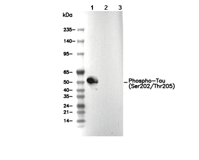 Lane 1: Mouse brain, Lane 2: Mouse brain (λ phosphatase-treated), Lane 3: Mouse brain (KO Tau)