|
Hoe te citeren 1. Voor in-tekst citatie (materialen en methoden): 2. Voor de tabel met belangrijke bronnen: |
||
|
Gratis nummer: (877) 796-6397 -- Alleen VS en Canada -- |
Fax: +1-832-582-8590 Bestellingen: +1-832-582-8158 |
Technische ondersteuning: +1-832-582-8158 Ext:3 Vermeld uw bestelnummer in de e-mail. Wij streven ernaar alle e-mailvragen binnen één werkdag te beantwoorden. |
Biologische Beschrijving
| Specificiteit | Phospho-Synapsin-1 (Ser605) Antibody [M6M7] detecteert endogene niveaus van Synapsin-1-eiwit alleen wanneer gefosforyleerd op Ser605 (komt overeen met Ser603 bij rat). |
|---|---|
| Achtergrond | Synapsin I (eiwit I) is een belangrijk neuron-specifiek fosfoproteïne en een cruciaal endogeen substraat voor zowel cAMP-afhankelijke als Ca²⁺/calmoduline-afhankelijke proteïnekinasen. Het is wijd verspreid in synapsen door het centrale en perifere zenuwstelsel, waar het specifiek geassocieerd is met het cytoplasmatische oppervlak van synaptische vesikelmembranen. De synapsine-eiwitfamilie bestaat uit vier homologe leden: synapsinen Ia en Ib (gezamenlijk aangeduid als synapsine I) en synapsinen IIa en IIb (gezamenlijk aangeduid als synapsine II). Samen zijn synapsinen I en II goed voor ongeveer 9% van het totale synaptische vesikeleiwit. Synapsinen I en II zijn voornamelijk aanwezig in volwassen synapsen, terwijl synapsine III voornamelijk tot expressie komt tijdens synapsontwikkeling en op relatief lagere niveaus. Functioneel speelt synapsine I een cruciale rol bij het reguleren van neurotransmitterafgifte. In neuronen helpt het de beschikbaarheid van synaptische vesikels voor exocytose te controleren. Fosforylering op het Ser-9-residu (fosfo-Ser9 synapsine I) zorgt ervoor dat het eiwit dissocieert van synaptische vesikels, een proces dat essentieel is voor neurotransmitterafgifte. Bovendien draagt synapsine I bij aan synaptische plasticiteit door zowel de pre- als postsynaptische vesikelafgifte te beïnvloeden. Genetisch zijn mutaties in het synapsine I-gen gekoppeld aan X-gebonden epilepsie met variabele leerstoornissen en gedragsstoornissen (XELBD), een neurologische aandoening die wordt gekenmerkt door variërende combinaties van epilepsie, cognitieve stoornissen, macrocefalie en agressief gedrag. Ser605 is bevestigd als een belangrijke fosforyleringsplaats op Synapsin I, met in vivo bewijs dat het belang ervan ondersteunt. Fosforylering op Ser605 (naast Ser568) door Ca²⁺/calmoduline-afhankelijke proteïnekinase II (CaMKII) verstoort het vermogen van Synapsin I om actinefilamenten te bundelen. Dit is waarschijnlijk een mechanisme dat de dynamische organisatie van het presynaptische cytoskelet reguleert. |
Gebruiksinformatie
| Toepassing | WB | Verdunning |
|
||
|---|---|---|---|---|---|
| Reactiviteit | Human, Mouse, Rat | ||||
| Bron | Rabbit Monoclonal Antibody | MW | 75-90 kDa | ||
| Opslagbuffer | PBS, pH 7.2+50% Glycerol+0.05% BSA+0.01% NaN3 | Opslag (Vanaf de datum van ontvangst) |
-20°C (avoid freeze-thaw cycles), 2 years | ||
| WB |
Experimental Protocol:
Sample preparation
1. Tissue: Lyse the tissue sample by adding an appropriate volume of ice-cold RIPA/NP-40 Lysis Buffer (containing Protease Inhibitor Cocktail, Phosphatase Inhibitor Cocktail),and homogenize the tissue at a low temperature. 2. Adherent cell: Aspirate the culture medium and wash the cells with ice-cold PBS twice. Lyse the cells by adding an appropriate volume of RIPA/NP-40 Lysis Buffer (containing Protease Inhibitor Cocktail, Phosphatase Inhibitor Cocktail) and put the sample on ice for 5 min. 3. Suspension cell: Transfer the culture medium to a pre-cooled centrifuge tube. Centrifuge and aspirate the supernatant. Wash the cells with ice-cold PBS twice. Lyse the cells by adding an appropriate volume of RIPA/NP-40 Lysis Buffer (containing Protease Inhibitor Cocktail, Phosphatase Inhibitor Cocktail) and put the sample on ice for 5 min. 4. Place the lysate into a pre-cooled microcentrifuge tube. Centrifuge at 4°C for 15 min. Collect the supernatant;
5. Remove a small volume of lysate to determine the protein concentration;
6. Combine the lysate with protein loading buffer. Boil 20 µL sample under 95-100°C for 5 min. Centrifuge for 5 min after cool down on ice.
Electrophoretic separation
1. According to the concentration of extracted protein, load appropriate amount of protein sample and marker onto SDS-PAGE gels for electrophoresis. Recommended separating gel (lower gel) concentration: 10%. Reference Table for Selecting SDS-PAGE Separation Gel Concentrations 2. Power up 80V for 30 minutes. Then the power supply is adjusted (110 V~150 V), the Marker is observed, and the electrophoresis can be stopped when the indicator band of the predyed protein Marker where the protein is located is properly separated. (Note that the current should not be too large when electrophoresis, too large current (more than 150 mA) will cause the temperature to rise, affecting the result of running glue. If high currents cannot be avoided, an ice bath can be used to cool the bath.)
Transfer membrane
1. Take out the converter, soak the clip and consumables in the pre-cooled converter;
2. Activate PVDF membrane with methanol for 1 min and rinse with transfer buffer;
3. Install it in the order of "black edge of clip - sponge - filter paper - filter paper - glue -PVDF membrane - filter paper - filter paper - sponge - white edge of clip"; 4. The protein was electrotransferred to PVDF membrane. ( 0.45 µm PVDF membrane is recommended ) Reference Table for Selecting PVDF Membrane Pore Size Specifications Recommended conditions for wet transfer: 200 mA, 120 min. ( Note that the transfer conditions can be adjusted according to the protein size. For high-molecular-weight proteins, a higher current and longer transfer time are recommended. However, ensure that the transfer tank remains at a low temperature to prevent gel melting.)
Block
1. After electrotransfer, wash the film with TBST at room temperature for 5 minutes;
2. Incubate the film in the blocking solution ( recommending 5% BSA solution)
for 1 hour at room temperature;
3. Wash the film with TBST for 3 times, 5 minutes each time.
Antibody incubation
1. Use 5% skim milk powder to prepare the primary antibody working liquid (recommended dilution ratio for primary antibody 1:1000), gently shake and incubate with the film at 4°C overnight; 2. Wash the film with TBST 3 times, 5 minutes each time;
3. Add the secondary antibody to the blocking solution and incubate with the film gently at room temperature for 1 hour;
4. After incubation, wash the film with TBST 3 times for 5 minutes each time.
Antibody staining
1. Add the prepared ECL luminescent substrate (or select other color developing substrate according to the second antibody) and mix evenly;
2. Incubate with the film for 1 minute, remove excess substrate (keep the film moist), wrap with plastic film, and expose in the imaging system. (Exposure time of at least 150s is recommended)
|
Referenties
|
Toepassingsgegevens
WB
Gevalideerd door Selleck
-
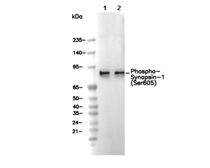 Lane 1: Mouse brain, Lane 2: Rat brain