|
Hoe te citeren 1. Voor in-tekst citatie (materialen en methoden): 2. Voor de tabel met belangrijke bronnen: |
||
|
Gratis nummer: (877) 796-6397 -- Alleen VS en Canada -- |
Fax: +1-832-582-8590 Bestellingen: +1-832-582-8158 |
Technische ondersteuning: +1-832-582-8158 Ext:3 Vermeld uw bestelnummer in de e-mail. Wij streven ernaar alle e-mailvragen binnen één werkdag te beantwoorden. |
Biologische Beschrijving
| Specificiteit | Phospho-Phospholipase Cγ1/PLC-γ-1 (Tyr783) Antibody [J4J15] detecteert endogene niveaus van totaal Phospholipase Cγ1/PLC-γ-1 eiwit alleen wanneer het gefosforyleerd is op Tyr783. |
|---|---|
| Achtergrond | Phospho-Phospholipase Cγ1 (Tyr783), de geactiveerde vorm van PLC-γ1, is een cruciale effector in de receptor tyrosine kinase (RTK) signalering. Bij RTK-stimulatie wordt Tyr783 van PLC-γ1 gefosforyleerd, typisch door kinasen zoals EGFR, PDGFR, Syk of leden van de Src-familie. Deze fosforyleringsgebeurtenis triggert een conformationele verandering, waardoor de autoinhibitie wordt opgeheven die wordt opgelegd door de gesplitste SH2-SH2-SH3-domeinen die anders de katalytische kern vastklemmen. De gefosforyleerde Tyr783 in de SH2-linker bindt intramoleculair aan het cSH2-domein, waardoor het wordt verplaatst van het C2-kerninterface en de actieve site wordt blootgesteld voor membraandocking en katalyse. PLC-γ1 kenmerkt een N-terminaal PH-domein voor PIP2-membraanrecruitment, een katalytische TIM-barrel (X-Y-domeinen), EF-handen en een C2-domein voor Ca2+/lipide detectie, en regulerende SH2-SH2-SH3-domeinen. Eenmaal geactiveerd, hydrolyseert PLC-γ1 fosfatidylinositol 4,5-bisfosfaat (PIP2) in inositol 1,4,5-trisfosfaat (IP3) en diacylglycerol (DAG). Deze second messengers drijven intracellulaire calciumafgifte en proteïne kinase C (PKC) activering, waardoor downstream signalering wordt voortgeplant. De hoge-affiniteit cSH2-pTyr783 interactie overtreft autoinhiberende contacten, stabiliseert een open, katalytisch actieve conformatie en versterkt de IP3/DAG-productie. Dit ondersteunt Ca2+-oscillaties, PKC/ERK-crosstalk, cytoskeletale remodeling, genexpressie en chemotaxis, vooral in immuuncellen. Snelle defosforylering van Tyr783 zorgt voor een tijdige signaalbeëindiging. Phospho-PLCγ1 is onmisbaar voor bloedplaatjesaggregatie, T- en B-celactivering en RTK-gedreven celproliferatie. Abberrante Tyr783-fosforylering kan immuunstoornissen (bijv. SCID door gain-of-function mutaties), hematologische maligniteiten via hyperactieve PLC-signalering veroorzaken en invasie en angiogenese in solide tumoren stimuleren. |
Gebruiksinformatie
| Toepassing | WB, IF | Verdunning |
|
||||
|---|---|---|---|---|---|---|---|
| Reactiviteit | Mouse, Human | ||||||
| Bron | Rabbit Monoclonal Antibody | MW | 148 kDa | ||||
| Opslagbuffer | PBS, pH 7.2+50% Glycerol+0.05% BSA+0.01% NaN3 | Opslag (Vanaf de datum van ontvangst) |
-20°C (avoid freeze-thaw cycles), 2 years | ||||
| WB |
Experimental Protocol:
Sample preparation
1. Tissue: Lyse the tissue sample by adding an appropriate volume of ice-cold RIPA/NP-40 Lysis Buffer (containing Protease Inhibitor Cocktail, Phosphatase Inhibitor Cocktail),and homogenize the tissue at a low temperature. 2. Adherent cell: Aspirate the culture medium and wash the cells with ice-cold PBS twice. Lyse the cells by adding an appropriate volume of RIPA/NP-40 Lysis Buffer (containing Protease Inhibitor Cocktail, Phosphatase Inhibitor Cocktail) and put the sample on ice for 5 min. 3. Suspension cell: Transfer the culture medium to a pre-cooled centrifuge tube. Centrifuge and aspirate the supernatant. Wash the cells with ice-cold PBS twice. Lyse the cells by adding an appropriate volume of RIPA/NP-40 Lysis Buffer (containing Protease Inhibitor Cocktail, Phosphatase Inhibitor Cocktail) and put the sample on ice for 5 min. 4. Place the lysate into a pre-cooled microcentrifuge tube. Centrifuge at 4°C for 15 min. Collect the supernatant;
5. Remove a small volume of lysate to determine the protein concentration;
6. Combine the lysate with protein loading buffer. Boil 20 µL sample under 95-100°C for 5 min. Centrifuge for 5 min after cool down on ice.
Electrophoretic separation
1. According to the concentration of extracted protein, load appropriate amount of protein sample and marker onto SDS-PAGE gels for electrophoresis. Recommended separating gel (lower gel) concentration: 5%. Reference Table for Selecting SDS-PAGE Separation Gel Concentrations 2. Power up 80V for 30 minutes. Then the power supply is adjusted (110 V~150 V), the Marker is observed, and the electrophoresis can be stopped when the indicator band of the predyed protein Marker where the protein is located is properly separated. (Note that the current should not be too large when electrophoresis, too large current (more than 150 mA) will cause the temperature to rise, affecting the result of running glue. If high currents cannot be avoided, an ice bath can be used to cool the bath.)
Transfer membrane
1. Take out the converter, soak the clip and consumables in the pre-cooled converter;
2. Activate PVDF membrane with methanol for 1 min and rinse with transfer buffer;
3. Install it in the order of "black edge of clip - sponge - filter paper - filter paper - glue -PVDF membrane - filter paper - filter paper - sponge - white edge of clip"; 4. The protein was electrotransferred to PVDF membrane. ( 0.45 µm PVDF membrane is recommended ) Reference Table for Selecting PVDF Membrane Pore Size Specifications Recommended conditions for wet transfer: 200 mA, 120 min. ( Note that the transfer conditions can be adjusted according to the protein size. For high-molecular-weight proteins, a higher current and longer transfer time are recommended. However, ensure that the transfer tank remains at a low temperature to prevent gel melting.)
Block
1. After electrotransfer, wash the film with TBST at room temperature for 5 minutes;
2. Incubate the film in the blocking solution ( recommending 5% BSA solution)
for 1 hour at room temperature;
3. Wash the film with TBST for 3 times, 5 minutes each time.
Antibody incubation
1. Use 5% skim milk powder to prepare the primary antibody working liquid (recommended dilution ratio for primary antibody 1:100000), gently shake and incubate with the film at 4°C overnight; 2. Wash the film with TBST 3 times, 5 minutes each time;
3. Add the secondary antibody to the blocking solution and incubate with the film gently at room temperature for 1 hour;
4. After incubation, wash the film with TBST 3 times for 5 minutes each time.
Antibody staining
1. Add the prepared ECL luminescent substrate (or select other color developing substrate according to the second antibody) and mix evenly;
2. Incubate with the film for 1 minute, remove excess substrate (keep the film moist), wrap with plastic film, and expose in the imaging system.
|
| IF |
Experimental Protocol:
Sample Preparation
1. Adherent Cells: Place a clean, sterile coverslip in a culture dish. Once the cells grow to near confluence as a monolayer, remove the coverslip for further use.
2. Suspension Cells: Seed the cells onto a clean, sterile slide coated with poly-L-lysine.
3. Frozen Sections: Allow the slide to thaw at room temperature. Wash it with pure water or PBS for 2 times, 3 minutes each time.
4. Paraffin Sections: Deparaffinization and rehydration. Wash the slide with pure water or PBS for 3 times, 3 minutes each time. Then perform antigen retrieval.
Fixation
1. Fix the cell coverslips/spots or tissue sections at room temperature using a fixative such as 4% paraformaldehyde (4% PFA) for 10-15 minutes.
2. Wash the sample with PBS for 3 times, 3 minutes each time.
Permeabilization
1.Add a detergent such as 0.1–0.3% Triton X-100 to the sample and incubate at room temperature for 10–20 minutes.
(Note: This step is only required for intracellular antigens. For antigens expressed on the cell membrane, this step is unnecessary.)
Wash the sample with PBS for 3 times, 3 minutes each time.
Blocking
Add blocking solution and incubate at room temperature for at least 1 hour. (Common blocking solutions include: serum from the same source as the secondary antibody, BSA, or goat serum.)
Note: Ensure the sample remains moist during and after the blocking step to prevent drying, which can lead to high background.
Immunofluorescence Staining (Day 1)
1. Remove the blocking solution and add the diluted primary antibody.
2. Incubate the sample in a humidified chamber at 4°C overnight.
Immunofluorescence Staining (Day 2)
1. Remove the primary antibody and wash with PBST for 3 times, 5 minutes each time.
2. Add the diluted fluorescent secondary antibody and incubate in the dark at 4°C for 1–2 hours.
3. Remove the secondary antibody and wash with PBST for 3 times, 5 minutes each time.
4. Add diluted DAPI and incubate at room temperature in the dark for 5–10 minutes.
5. Wash with PBST for 3 times, 5 minutes each time.
Mounting
1. Mount the sample with an anti-fade mounting medium.
2. Allow the slide to dry at room temperature overnight in the dark.
3. Store the slide in a slide storage box at 4°C, protected from light.
|
Referenties
|
Toepassingsgegevens
WB
Gevalideerd door Selleck
-
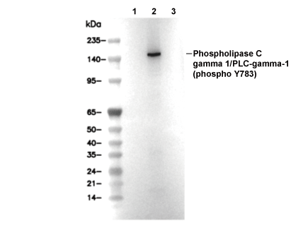 Lane 1: Jurkat, Lane 2: Jurkat (pervanadate, 50mM, 5 min), Lane 3: Jurkat (pervanadate, 50mM, 5 min; alkaline phosphatase treated)