|
Hoe te citeren 1. Voor in-tekst citatie (materialen en methoden): 2. Voor de tabel met belangrijke bronnen: |
||
|
Gratis nummer: (877) 796-6397 -- Alleen VS en Canada -- |
Fax: +1-832-582-8590 Bestellingen: +1-832-582-8158 |
Technische ondersteuning: +1-832-582-8158 Ext:3 Vermeld uw bestelnummer in de e-mail. Wij streven ernaar alle e-mailvragen binnen één werkdag te beantwoorden. |
Biologische Beschrijving
| Specificiteit | Phospho-NF-κB p65 (Ser468) Antibody [G12A6] herkent endogene niveaus van totaal NF-κB p65-eiwit alleen wanneer gefosforyleerd op Ser468. |
|---|---|
| Achtergrond | Phospho-NF-κB p65 (Ser468) verwijst naar de fosforylering van de p65 (RelA)-subeenheid van de NF-κB-familie op serine 468 binnen het C-terminale transactivatiedomein. De NF-κB-familie reguleert genen die betrokken zijn bij immuniteit, ontsteking en celoverleving via dimere transcriptiefactorcomplexen. p65 bevat een Rel-homologiedomein dat verantwoordelijk is voor DNA-binding en dimerisatie, en een transactivatiedomein dat transcriptionele activering medieert. Fosforylering op Ser468 wordt gemedieerd door IKKε tijdens T-cel co-stimulatie en door GSK-3β onder basale omstandigheden. Deze post-translationele modificatie bevordert de binding van p65 aan het COMMD1–Cullin2 ubiquitine-ligasecomplex, wat leidt tot ubiquitinatie en proteasomale afbraak van chromatine-gebonden p65. Fosforylering op Ser468 beëindigt selectief de transcriptie van specifieke NF-κB target genes, zoals ICAM1, door het verwijderen van p65 uit chromatine te vergemakkelijken. Mutatie van Ser468 verstoort dit regulerende mechanisme, stabiliseert p65 en verlengt de transcriptionele activiteit ervan. GSK-3β-gemedieerde fosforylering onderdrukt basale NF-κB-signalering, terwijl IKKε-gedreven fosforylering de reacties op immuunstimuli verfijnt. Deze modificatie dient dus als een integratiepunt voor constitutieve en induceerbare regulerende signalen, waardoor een precieze controle van NF-κB-activiteit mogelijk wordt. Dysregulatie van Ser468-fosforylering wordt geassocieerd met chronische ontsteking, auto-immuniteit en tumorigenese. |
Gebruiksinformatie
| Toepassing | WB, FCM, ELISA | Verdunning |
|
||
|---|---|---|---|---|---|
| Reactiviteit | Human, Mouse | ||||
| Bron | Rabbit Monoclonal Antibody | MW | 65 kDa | ||
| Opslagbuffer | PBS, pH 7.2+50% Glycerol+0.05% BSA+0.01% NaN3 | Opslag (Vanaf de datum van ontvangst) |
-20°C (avoid freeze-thaw cycles), 2 years | ||
| WB |
Experimental Protocol:
Sample preparation
1. Tissue: Lyse the tissue sample by adding an appropriate volume of ice-cold RIPA/Nuclear Lysis Buffer (containing Protease Inhibitor Cocktail, Phosphatase Inhibitor Cocktail),and homogenize the tissue at a low temperature. 2. Adherent cell: Aspirate the culture medium and wash the cells with ice-cold PBS twice. Lyse the cells by adding an appropriate volume of RIPA/Nuclear Lysis Buffer (containing Protease Inhibitor Cocktail, Phosphatase Inhibitor Cocktail) and put the sample on ice for 5 min. 3. Suspension cell: Transfer the culture medium to a pre-cooled centrifuge tube. Centrifuge and aspirate the supernatant. Wash the cells with ice-cold PBS twice. Lyse the cells by adding an appropriate volume of RIPA/Nuclear Lysis Buffer (containing Protease Inhibitor Cocktail, Phosphatase Inhibitor Cocktail) and put the sample on ice for 5 min. 4. Place the lysate into a pre-cooled microcentrifuge tube. Centrifuge at 4°C for 15 min. Collect the supernatant;
5. Remove a small volume of lysate to determine the protein concentration;
6. Combine the lysate with protein loading buffer. Boil 20 µL sample under 95-100°C for 5 min. Centrifuge for 5 min after cool down on ice.
Electrophoretic separation
1. According to the concentration of extracted protein, load appropriate amount of protein sample and marker onto SDS-PAGE gels for electrophoresis. Recommended separating gel (lower gel) concentration: 10%. Reference Table for Selecting SDS-PAGE Separation Gel Concentrations 2. Power up 80V for 30 minutes. Then the power supply is adjusted (110 V~150 V), the Marker is observed, and the electrophoresis can be stopped when the indicator band of the predyed protein Marker where the protein is located is properly separated. (Note that the current should not be too large when electrophoresis, too large current (more than 150 mA) will cause the temperature to rise, affecting the result of running glue. If high currents cannot be avoided, an ice bath can be used to cool the bath.)
Transfer membrane
1. Take out the converter, soak the clip and consumables in the pre-cooled converter;
2. Activate PVDF membrane with methanol for 1 min and rinse with transfer buffer;
3. Install it in the order of "black edge of clip - sponge - filter paper - filter paper - glue -PVDF membrane - filter paper - filter paper - sponge - white edge of clip"; 4. The protein was electrotransferred to PVDF membrane. ( 0.45 µm PVDF membrane is recommended ) Reference Table for Selecting PVDF Membrane Pore Size Specifications Recommended conditions for wet transfer: 200 mA, 120 min. ( Note that the transfer conditions can be adjusted according to the protein size. For high-molecular-weight proteins, a higher current and longer transfer time are recommended. However, ensure that the transfer tank remains at a low temperature to prevent gel melting.)
Block
1. After electrotransfer, wash the film with TBST at room temperature for 5 minutes;
2. Incubate the film in the blocking solution ( recommending 5% BSA solution)
for 1 hour at room temperature;
3. Wash the film with TBST for 3 times, 5 minutes each time.
Antibody incubation
1. Use 5% skim milk powder to prepare the primary antibody working liquid (recommended dilution ratio for primary antibody 1:2000), gently shake and incubate with the film at 4°C overnight; 2. Wash the film with TBST 3 times, 5 minutes each time;
3. Add the secondary antibody to the blocking solution and incubate with the film gently at room temperature for 1 hour;
4. After incubation, wash the film with TBST 3 times for 5 minutes each time.
Antibody staining
1. Add the prepared ECL luminescent substrate (or select other color developing substrate according to the second antibody) and mix evenly;
2. Incubate with the film for 1 minute, remove excess substrate (keep the film moist), wrap with plastic film, and expose in the imaging system.
|
Referenties
|
Toepassingsgegevens
WB
Gevalideerd door Selleck
-
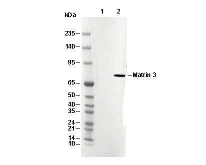 Lane 1: Hela, Lane 2: Hela (Calyculin A, 100nM, 30 min)