|
Hoe te citeren 1. Voor in-tekst citatie (materialen en methoden): 2. Voor de tabel met belangrijke bronnen: |
||
|
Gratis nummer: (877) 796-6397 -- Alleen VS en Canada -- |
Fax: +1-832-582-8590 Bestellingen: +1-832-582-8158 |
Technische ondersteuning: +1-832-582-8158 Ext:3 Vermeld uw bestelnummer in de e-mail. Wij streven ernaar alle e-mailvragen binnen één werkdag te beantwoorden. |
Biologische Beschrijving
| Specificiteit | Phospho-Hsp27 (Ser82) Antibody [M1P19] detecteert endogene niveaus van totaal Phospho-HSP27 (Ser82) eiwit alleen wanneer gefosforyleerd op Ser82. |
|---|---|
| Achtergrond | Hitte-shockproteïne 27 (HSP27) is een lid van de familie van kleine hitte-shockproteïnen (sHSP) (12–43 kDa) en vertoont een breed scala aan cellulaire functies. Het fungeert als een moleculaire chaperonne, een antioxidant, een regulator van apoptose en een mediator van actine cytoskeletale remodellering. Net als andere kleine HSP's bevat HSP27 een geconserveerd α-crystallinedomein in het C-terminale gebied. Onder oxidatieve stress beschermt HSP27 cellen door de intracellulaire niveaus van reactieve zuurstofsoorten (ROS) te verlagen, wat wordt bereikt door de glutathionproductie te verbeteren en de beschikbaarheid van vrij ijzer te verminderen. Het vertoont ook een sterke anti-apoptotische activiteit, die zowel mitochondriën-afhankelijke als onafhankelijke apoptotische routes beïnvloedt. HSP27 bindt bijvoorbeeld aan DAXX tijdens Fas–FasL–gemedieerde apoptose, waardoor DAXX niet kan associëren met ASK1. Bovendien interageert het met Bax en cytochroom c, waardoor mitochondriale apoptose wordt onderdrukt en caspase-afhankelijke celdood wordt geblokkeerd. HSP27 wordt op basale niveaus tot expressie gebracht in de meeste cellen en weefsels, waar het typisch bestaat als grote oligomere complexen. Bij blootstelling aan stress stijgt de expressie ervan significant, waardoor de cellulaire weerstand tegen schadelijke omstandigheden wordt verbeterd. Het eiwit is onderhevig aan stress-geïnduceerde fosforylering op Ser15, Ser78 en Ser82 bij mensen (Ser15 en Ser86 bij knaagdieren), gemedieerd door MAPKAP kinase 2/3 stroomafwaarts van de p38 MAPK signaalroute. Fosforylering beïnvloedt niet alleen de oligomere staat, maar reguleert ook de interacties met partnerproteïnen. In neutrofielen vormt HSP27 een complex met AKT en MAPKAP kinase 2, dat constitutieve apoptose onderdrukt en een ontstekingsreactie bevordert. Stress-geïnduceerde fosforylering van HSP27 verstoort dit complex, wat leidt tot dissociatie van AKT en herstel van neutrofiele apoptose. Met name zijn de effecten van gefosforyleerd HSP27 sterk contextafhankelijk, variërend met celtype en signaalomgeving. |
Gebruiksinformatie
| Toepassing | WB, IP | Verdunning |
|
||||
|---|---|---|---|---|---|---|---|
| Reactiviteit | Mouse, Rat, Human | ||||||
| Bron | Rabbit Monoclonal Antibody | MW | 23 kDa | ||||
| Opslagbuffer | PBS, pH 7.2+50% Glycerol+0.05% BSA+0.01% NaN3 | Opslag (Vanaf de datum van ontvangst) |
-20°C (avoid freeze-thaw cycles), 2 years | ||||
| WB |
Experimental Protocol:
Sample preparation
1. Tissue: Lyse the tissue sample by adding an appropriate volume of ice-cold RIPA/NP-40 Lysis Buffer (containing Protease Inhibitor Cocktail, Phosphatase Inhibitor Cocktail),and homogenize the tissue at a low temperature. 2. Adherent cell: Aspirate the culture medium and wash the cells with ice-cold PBS twice. Lyse the cells by adding an appropriate volume of RIPA/NP-40 Lysis Buffer (containing Protease Inhibitor Cocktail, Phosphatase Inhibitor Cocktail) and put the sample on ice for 5 min. 3. Suspension cell: Transfer the culture medium to a pre-cooled centrifuge tube. Centrifuge and aspirate the supernatant. Wash the cells with ice-cold PBS twice. Lyse the cells by adding an appropriate volume of RIPA/NP-40 Lysis Buffer (containing Protease Inhibitor Cocktail, Phosphatase Inhibitor Cocktail) and put the sample on ice for 5 min. 4. Place the lysate into a pre-cooled microcentrifuge tube. Centrifuge at 4°C for 15 min. Collect the supernatant;
5. Remove a small volume of lysate to determine the protein concentration;
6. Combine the lysate with protein loading buffer. Boil 20 µL sample under 95-100°C for 5 min. Centrifuge for 5 min after cool down on ice.
Electrophoretic separation
1. According to the concentration of extracted protein, load appropriate amount of protein sample and marker onto SDS-PAGE gels for electrophoresis. Recommended separating gel (lower gel) concentration: 10%. Reference Table for Selecting SDS-PAGE Separation Gel Concentrations 2. Power up 80V for 30 minutes. Then the power supply is adjusted (110 V~150 V), the Marker is observed, and the electrophoresis can be stopped when the indicator band of the predyed protein Marker where the protein is located is properly separated. (Note that the current should not be too large when electrophoresis, too large current (more than 150 mA) will cause the temperature to rise, affecting the result of running glue. If high currents cannot be avoided, an ice bath can be used to cool the bath.)
Transfer membrane
1. Take out the converter, soak the clip and consumables in the pre-cooled converter;
2. Activate PVDF membrane with methanol for 1 min and rinse with transfer buffer;
3. Install it in the order of "black edge of clip - sponge - filter paper - filter paper - glue -PVDF membrane - filter paper - filter paper - sponge - white edge of clip"; 4. The protein was electrotransferred to PVDF membrane. ( 0.45 µm PVDF membrane is recommended ) Reference Table for Selecting PVDF Membrane Pore Size Specifications Recommended conditions for wet transfer: 200 mA, 60 min. ( Note that the transfer conditions can be adjusted according to the protein size. For high-molecular-weight proteins, a higher current and longer transfer time are recommended. However, ensure that the transfer tank remains at a low temperature to prevent gel melting.)
Block
1. After electrotransfer, wash the film with TBST at room temperature for 5 minutes;
2. Incubate the film in the blocking solution ( recommending 5% BSA solution)
for 1 hour at room temperature;
3. Wash the film with TBST for 3 times, 5 minutes each time.
Antibody incubation
1. Use 5% skim milk powder to prepare the primary antibody working liquid (recommended dilution ratio for primary antibody 1:1000), gently shake and incubate with the film at 4°C overnight; 2. Wash the film with TBST 3 times, 5 minutes each time;
3. Add the secondary antibody to the blocking solution and incubate with the film gently at room temperature for 1 hour;
4. After incubation, wash the film with TBST 3 times for 5 minutes each time.
Antibody staining
1. Add the prepared ECL luminescent substrate (or select other color developing substrate according to the second antibody) and mix evenly;
2. Incubate with the film for 1 minute, remove excess substrate (keep the film moist), wrap with plastic film, and expose in the imaging system.
|
Referenties
|
Toepassingsgegevens
WB
Gevalideerd door Selleck
-
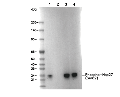 Lane 1: HeLa (with 44°C heat shock), Lane 2: HeLa, Lane 3: Mouse heart, Lane 4: Rat heart