|
Hoe te citeren 1. Voor in-tekst citatie (materialen en methoden): 2. Voor de tabel met belangrijke bronnen: |
||
|
Gratis nummer: (877) 796-6397 -- Alleen VS en Canada -- |
Fax: +1-832-582-8590 Bestellingen: +1-832-582-8158 |
Technische ondersteuning: +1-832-582-8158 Ext:3 Vermeld uw bestelnummer in de e-mail. Wij streven ernaar alle e-mailvragen binnen één werkdag te beantwoorden. |
Biologische Beschrijving
| Specificiteit | ID3 Antibody [J14L22] herkent endogene niveaus van totaal ID3-eiwit. |
|---|---|
| Achtergrond | ID3 (Inhibitor of DNA-binding 3) is een lid van de helix-loop-helix (HLH) familie van transcriptiefactoren en speelt een centrale rol bij het reguleren van belangrijke cellulaire processen zoals differentiatie, proliferatie en DNA Damage/DNA Repair (DDR). In tegenstelling tot andere HLH-eiwitten mist ID3 een basisch DNA-bindend domein en functioneert het in plaats daarvan door te binden aan en E-eiwitten, transcriptiefactoren die cellulaire differentiatie bevorderen, te sequesteren. Deze interactie voorkomt dat E-eiwitten genen activeren die geassocieerd zijn met differentiatie, waardoor een progenitor- of ongedifferentieerde staat behouden blijft. De structuur van ID3 omvat een basisch HLH-domein, cruciaal voor eiwit-eiwitinteracties maar niet voor directe DNA-binding. Bij DNA Damage/DNA Repair ondergaat ID3 fosforylering op serine 65 door het ATM (Ataxia Telangiectasia Mutated) kinase, een belangrijke regulator van de DDR. Deze fosforyleringsgebeurtenis is essentieel voor het verbeteren van de interacties met eiwitten die betrokken zijn bij DNA Damage/DNA Repair, met name die betrokken zijn bij de reparatie van dubbelstrengsbreuken (DSB's), zoals het MRN-complex (NBS1, RAD50, MRE11), MDC1 en RECQL. Deze interacties zijn cruciaal voor het vergemakkelijken van reparatie door homologe recombinatie (HR), waarbij ID3 de rekrutering van belangrijke reparatie-enzymen, zoals RAD51, bevordert en DNA-eindresectie vergemakkelijkt. Het helpt ook bij transcriptieregulatie, waar het de expressie van DNA Damage/DNA Repair-genen, zoals die betrokken zijn bij HR- en Fanconi Anemia (FA)-routes, beïnvloedt via interacties met transcriptiefactoren zoals E2F1 en chromatine-remodelleringsfactoren zoals PRMT5. Verlies van ID3 resulteert in verminderde DNA Damage/DNA Repair en verhoogde gevoeligheid voor DNA-beschadigende middelen, waaronder ioniserende straling en chemotherapeutische middelen zoals cisplatine. ID3 heeft een dubbele rol in de kankerbiologie, waar het zowel als tumorsuppressor als als promotor kan fungeren, afhankelijk van de context. Bij sommige kankers, zoals hematologische maligniteiten en colorectale kanker, bevordert ID3 de zelfvernieuwing van kankerstamcellen en chemotherapieresistentie, terwijl het bij andere de tumorprogressie onderdrukt door differentiatie te bevorderen en ongecontroleerde celproliferatie te voorkomen. |
Gebruiksinformatie
| Toepassing | WB, IP, IF, FCM | Verdunning |
|
||||||||
|---|---|---|---|---|---|---|---|---|---|---|---|
| Reactiviteit | Human | ||||||||||
| Bron | Rabbit Monoclonal Antibody | MW | 13 KDa | ||||||||
| Opslagbuffer | PBS, pH 7.2+50% Glycerol+0.05% BSA+0.01% NaN₃ | Opslag (Vanaf de datum van ontvangst) |
-20°C (avoid freeze-thaw cycles), 2 years | ||||||||
| WB |
Experimental Protocol:
Sample preparation
1. Tissue: Lyse the tissue sample by adding an appropriate volume of ice-cold RIPA/Nuclear Lysis Buffer (containing Protease Inhibitor Cocktail),and homogenize the tissue at a low temperature or lyse it by sonication on ice, then incubate on ice for 30 minutes. 2. Adherent cell: Aspirate the culture medium and transfer the cells into an EP tube. Wash the cells with ice-cold PBS twice. Add an appropriate volume of RIPA/Nuclear Lysis Buffer (containing Protease Inhibitor Cocktail), sonicate to lyse the cells, and incubate on ice for 30 minutes. 3. Suspension cell: Transfer the culture medium to a pre-cooled centrifuge tube. Centrifuge and aspirate the supernatant. Wash the cells with ice-cold PBS twice.Add an appropriate volume of RIPA/Nuclear Lysis Buffer (containing Protease Inhibitor Cocktail), sonicate to lyse the cells, and incubate on ice for 30 minutes. 4. Place the lysate into a pre-cooled microcentrifuge tube. Centrifuge at 4°C for 15 min. Collect the supernatant;
5. Remove a small volume of lysate to determine the protein concentration;
6. Combine the lysate with protein loading buffer. Boil 20 µL sample under 95-100°C for 5 min. Centrifuge for 5 min after cool down on ice.
Electrophoretic separation
1. According to the concentration of extracted protein, load appropriate amount of protein sample and marker onto SDS-PAGE gels for electrophoresis. Recommended separating gel (lower gel) concentration: 20%. Reference Table for Selecting SDS-PAGE Separation Gel Concentrations 2. Power up 80V for 30 minutes. Then the power supply is adjusted (110 V~150 V), the Marker is observed, and the electrophoresis can be stopped when the indicator band of the predyed protein Marker where the protein is located is properly separated. (Note that the current should not be too large when electrophoresis, too large current (more than 150 mA) will cause the temperature to rise, affecting the result of running glue. If high currents cannot be avoided, an ice bath can be used to cool the bath.)
Transfer membrane
1. Take out the converter, soak the clip and consumables in the pre-cooled converter;
2. Activate PVDF membrane with methanol for 1 min and rinse with transfer buffer;
3. Install it in the order of "black edge of clip - sponge - filter paper - filter paper - glue -PVDF membrane - filter paper - filter paper - sponge - white edge of clip"; 4. The protein was electrotransferred to PVDF membrane. ( 0.22 µm PVDF membrane is recommended )) Reference Table for Selecting PVDF Membrane Pore Size Specifications Recommended conditions for wet transfer: 200 mA, 60 min. ( Note that the transfer conditions can be adjusted according to the protein size. For high-molecular-weight proteins, a higher current and longer transfer time are recommended. However, ensure that the transfer tank remains at a low temperature to prevent gel melting.)
Block
1. After electrotransfer, wash the film with TBST at room temperature for 5 minutes;
2. Incubate the film in the blocking solution for 1 hour at room temperature;
3. Wash the film with TBST for 3 times, 5 minutes each time.
Antibody incubation
1. Use 5% skim milk powder to prepare the primary antibody working liquid (recommended dilution ratio for primary antibody 1:1000), gently shake and incubate with the film at 4°C overnight; 2. Wash the film with TBST 3 times, 5 minutes each time;
3. Add the secondary antibody to the blocking solution and incubate with the film gently at room temperature for 1 hour;
4. After incubation, wash the film with TBST 3 times for 5 minutes each time.
Antibody staining
1223. Add the prepared ECL luminescent substrate (or select other color developing substrate according to the second antibody) and mix evenly;
2. Incubate with the film for 1 minute, remove excess substrate (keep the film moist), wrap with plastic film, and expose in the imaging system. (Exposure time of at least 300s is recommended)
|
| IF |
Experimental Protocol:
Sample Preparation
1. Adherent Cells: Place a clean, sterile coverslip in a culture dish. Once the cells grow to near confluence as a monolayer, remove the coverslip for further use.
2. Suspension Cells: Seed the cells onto a clean, sterile slide coated with poly-L-lysine.
3. Frozen Sections: Allow the slide to thaw at room temperature. Wash it with pure water or PBS for 2 times, 3 minutes each time.
4. Paraffin Sections: Deparaffinization and rehydration. Wash the slide with pure water or PBS for 3 times, 3 minutes each time. Then perform antigen retrieval.
Fixation
1. Fix the cell coverslips/spots or tissue sections at room temperature using a fixative such as 4% paraformaldehyde (4% PFA) for 10-15 minutes.
2. Wash the sample with PBS for 3 times, 3 minutes each time.
Permeabilization
1.Add a detergent such as 0.1–0.3% Triton X-100 to the sample and incubate at room temperature for 10–20 minutes.
(Note: This step is only required for intracellular antigens. For antigens expressed on the cell membrane, this step is unnecessary.)
Wash the sample with PBS for 3 times, 3 minutes each time.
Blocking
Add blocking solution and incubate at room temperature for at least 1 hour. (Common blocking solutions include: serum from the same source as the secondary antibody, BSA, or goat serum.)
Note: Ensure the sample remains moist during and after the blocking step to prevent drying, which can lead to high background.
Immunofluorescence Staining (Day 1)
1. Remove the blocking solution and add the diluted primary antibody.
2. Incubate the sample in a humidified chamber at 4°C overnight.
Immunofluorescence Staining (Day 2)
1. Remove the primary antibody and wash with PBST for 3 times, 5 minutes each time.
2. Add the diluted fluorescent secondary antibody and incubate in the dark at 4°C for 1–2 hours.
3. Remove the secondary antibody and wash with PBST for 3 times, 5 minutes each time.
4. Add diluted DAPI and incubate at room temperature in the dark for 5–10 minutes.
5. Wash with PBST for 3 times, 5 minutes each time.
Mounting
1. Mount the sample with an anti-fade mounting medium.
2. Allow the slide to dry at room temperature overnight in the dark.
3. Store the slide in a slide storage box at 4°C, protected from light.
|
Referenties
|
Toepassingsgegevens
WB
Gevalideerd door Selleck
-
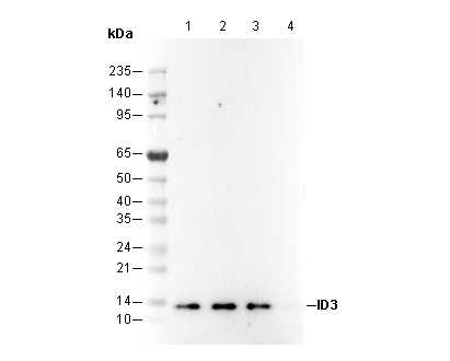 Lane 1: Hela
Lane 2: Jurkat
Lane 3: Ramos
IF
Gevalideerd door Selleck
-
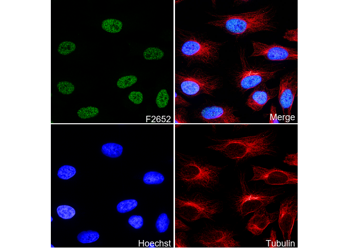 Immunofluorescent analysis of Hela cells using F2652 (green, 1:400), Hoechst (blue) and tubulin (Red).