Technische gegevens
| Formule | C25H17F2N5O3S |
||||||
| Moleculair gewicht | 505.5 | CAS-nr. | 1086062-66-9 | ||||
| Oplosbaarheid (25°C)* | In vitro | DMSO | 101 mg/mL (199.8 mM) | ||||
| Water | Insoluble | ||||||
| Ethanol | Insoluble | ||||||
| In vivo (Voeg oplosmiddelen afzonderlijk en in volgorde toe aan het product.) |
|
||||||
|
* <1 mg/ml betekent licht oplosbaar of onoplosbaar. * Houd er rekening mee dat Selleck de oplosbaarheid van alle verbindingen intern test en de werkelijke oplosbaarheid enigszins kan afwijken van gepubliceerde waarden. Dit is normaal en is te wijten aan lichte batch-tot-batch variaties. * Verzending op kamertemperatuur (Stabiliteitstests tonen aan dat dit product zonder koelmaatregelen kan worden verzonden.) |
|||||||
Voorbereiden van stamoplossingen
Biologische activiteit
| Beschrijving | Omipalisib (GSK2126458, GSK458) is een zeer selectieve en potente remmer van p110α/β/δ/γ, mTORC1/2 met een Ki van respectievelijk 0,019 nM/0,13 nM/0,024 nM/0,06 nM en 0,18 nM/0,3 nM in celvrije assays. Deze verbinding induceert Autophagy. Fase 1. | |||||||||||
|---|---|---|---|---|---|---|---|---|---|---|---|---|
| Doelen |
|
|||||||||||
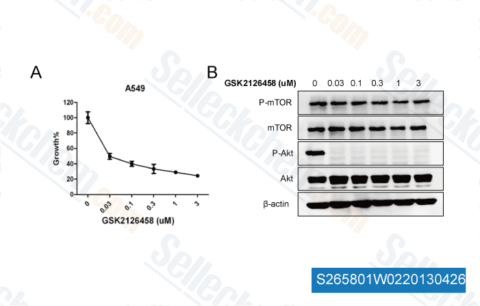
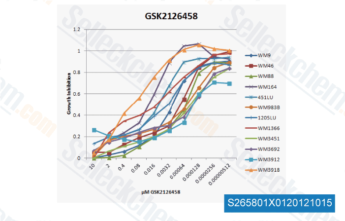
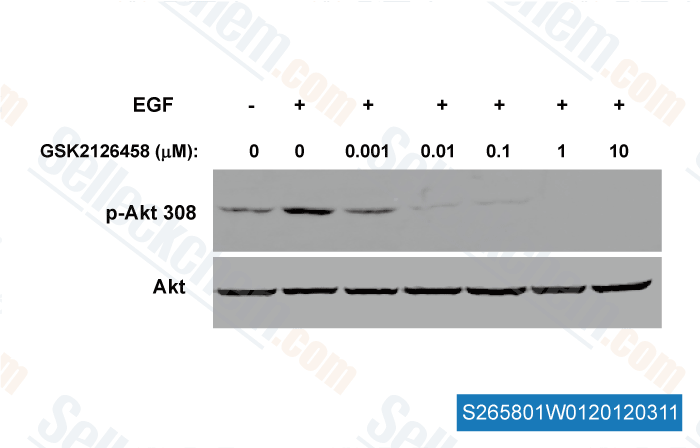
| In vitro | Omipalisib (GSK2126458) remt potent de activiteit van veelvoorkomende activerende mutanten van p110α (E542K, E545K en H1047R) die in menselijke kanker voorkomen, met een Ki van respectievelijk 8 pM, 8 pM en 9 pM. Deze verbinding veroorzaakt een significante reductie van de pAkt-S473-niveaus met opmerkelijke potentie in T47D- en BT474-cellen met een IC50 van respectievelijk 0,41 nM en 0,18 nM. Bovendien leidt het tot een G1-celcyclusarrest en produceert het een remmend effect op celproliferatie in een groot panel van cellijnen, waaronder de T47D- en BT474-borstkankerlijnen met een IC50 van respectievelijk 3 nM en 2,4 nM. | |||||||||||
| In vivo | In een BT474 humaan tumoxenograftmodel resulteert Omipalisib (GSK2126458) behandeling in een dosisafhankelijke reductie van pAkt-S473-niveaus, en vertoonde dosisafhankelijke tumorgroeiremming bij een lage dosis van 300 μg/kg. Bovendien vertoont het een lage bloedklaring en een goede orale biologische beschikbaarheid in vier preklinische soorten (muis, rat, hond en aap). |
Protocol (uit referentie)
| Kinase-assay:[1] |
|
|---|---|
| Celassay:[1] |
|
| Dierstudie:[1] |
|
Referenties
|
Klantproductvalidatie
-
Dr.Wang Wei from NanFang Hospital,
-
, , One customer
-
, , Dr. Zhang of Tianjin Medical University
-
Gegevens van [ , , Cancer Lett, 2017, 406:47-53 ]
Sellecks Omipalisib (GSK2126458) Is geciteerd door 61 Publicaties
| Depleting the action of EZH2 through PI3K-mTOR inhibition to overcome metastasis and immunotherapy resistance in triple-negative breast cancer [ Mol Cancer Ther, 2025, 10.1158/1535-7163.MCT-24-0693] | PubMed: 40497697 |
| Changes in Melanoma Cell Morphology Following Inhibition of Cell Invasion by Third-Generation mTOR Kinase Inhibitors [ Int J Mol Sci, 2025, 26(16)7770] | PubMed: 40869090 |
| Combined Omipalisib and MAPK Inhibition Suppress PDAC Growth [ Cancers (Basel), 2025, 17(7)1152] | PubMed: 40227649 |
| Trametinib sensitizes KRAS-mutant lung adenocarcinoma tumors to PD-1/PD-L1 axis blockade via Id1 downregulation [ Mol Cancer, 2024, 23(1):78] | PubMed: 38643157 |
| NKX2-5 regulates vessel remodeling in scleroderma-associated pulmonary arterial hypertension [ JCI Insight, 2024, 9(10)e164191] | PubMed: 38652537 |
| Characterizing heterogeneous single-cell dose responses computationally and experimentally using threshold inhibition surfaces and dose-titration assays [ NPJ Syst Biol Appl, 2024, 10(1):42] | PubMed: 38637530 |
| Analysis of hsa_circ_0136256 as a biomarker for fibrosis in systemic sclerosis [ BMC Biotechnol, 2024, 24(1):91] | PubMed: 39538329 |
| "Proteotranscriptomic analysis of advanced colorectal cancer patient derived organoids for drug sensitivity prediction" [ J Exp Clin Cancer Res, 2023, 42(1):8] | PubMed: 36604765 |
| PI3K/mTOR inhibitors promote G6PD autophagic degradation and exacerbate oxidative stress damage to radiosensitize small cell lung cancer [ Cell Death Dis, 2023, 14(10):652] | PubMed: 37802999 |
| Integrin-mediated electric axon guidance underlying optic nerve formation in the embryonic chick retina [ Commun Biol, 2023, 6(1):680] | PubMed: 37391492 |
RETOURBELEID
Selleck Chemicals onvoorwaardelijke retourbeleid zorgt voor een soepele online winkelervaring voor onze klanten. Als u op enigerlei wijze ontevreden bent met uw aankoop, kunt u elk artikel(en) binnen 7 dagen na ontvangst retourneren. In geval van problemen met de productkwaliteit, zowel protocolgerelateerde als productgerelateerde problemen, kunt u elk artikel(en) binnen 365 dagen na de oorspronkelijke aankoopdatum retourneren. Volg de onderstaande instructies bij het retourneren van producten.
VERZENDING EN OPSLAG
Selleck producten worden bij kamertemperatuur vervoerd. Als u het product op kamertemperatuur ontvangt, wees dan gerust, de Selleck kwaliteitsinspectieafdeling heeft experimenten uitgevoerd om te controleren of de normale temperatuurplaatsing van één maand de biologische activiteit van poederproducten niet beïnvloedt. Na ontvangst dient u het product op te slaan volgens de vereisten beschreven in het gegevensblad. De meeste Selleck producten zijn stabiel onder de aanbevolen omstandigheden.
NIET VOOR HUMANE, VETERINAIRE DIAGNOSTISCHE OF THERAPEUTISCHE DOELEINDEN.