|
Hoe te citeren 1. Voor in-tekst citatie (materialen en methoden): 2. Voor de tabel met belangrijke bronnen: |
||
|
Gratis nummer: (877) 796-6397 -- Alleen VS en Canada -- |
Fax: +1-832-582-8590 Bestellingen: +1-832-582-8158 |
Technische ondersteuning: +1-832-582-8158 Ext:3 Vermeld uw bestelnummer in de e-mail. Wij streven ernaar alle e-mailvragen binnen één werkdag te beantwoorden. |
Biologische Beschrijving
| Specificiteit | Glutathione Antibody [A11G7] detecteert endogene niveaus van totaal Glutathione-eiwit. |
|---|---|
| Achtergrond | Glutathione (GSH) is een tripeptide thiol-antioxidant met een laag molecuulgewicht, samengesteld uit glutamaat, cysteïne en glycine, gesynthetiseerd via twee opeenvolgende ATP-afhankelijke stappen, gekatalyseerd door glutamaat-cysteïne ligase en glutathion synthetase. Het bestaat voornamelijk in gereduceerde (GSH) en geoxideerde disulfide (GSSG) vormen, die onderling omgezet worden om de cellulaire redoxbalans te reguleren. GSH heeft een unieke γ-glutamyl peptidebinding tussen de γ-carboxylgroep van glutamaat en de aminogroep van cysteïne, wat resistentie verleent tegen afbraak door γ-glutamyl cyclotransferase en de meeste peptidasen; de cysteïne thiol (-SH) fungeert als het nucleofiele centrum voor redoxchemie, terwijl glycine de C-terminus stabiliseert. GSH vangt direct reactieve zuurstof- en stikstofspecies (ROS/RNS) weg via thiol-disulfide-uitwisseling, dient als een essentieel reductiemiddel voor glutathionperoxidasen (GPx) die peroxiden ontgiften (waarbij GSSG door glutathionreductase met behulp van NADPH wordt teruggereduceerd tot GSH), en conjugaten elektrofiele xenobiotica, zware metalen en endogene toxines via glutathion S-transferasen (GSTs) voor fase II-detoxificatie en mercaptuurzuur-export. Deze processen handhaven de proteïne-sulfhydrylhomeostase, ondersteunen de Nrf2-gedreven antioxidant-genexpressie, vergemakkelijken de assemblage van ijzer-zwavelclusters en metaaltransport, en moduleren signaalroutes zoals NF-κB. GSH-depletie is betrokken bij oxidatieve stressgerelateerde ziekten, waaronder de ziekte van Parkinson, levercirrose, kanker, diabetes en vroegtijdige veroudering. |
Gebruiksinformatie
| Toepassing | IF, FCM | Verdunning |
|
||||
|---|---|---|---|---|---|---|---|
| Reactiviteit | |||||||
| Bron | Mouse Monoclonal Antibody | MW | |||||
| Opslagbuffer | PBS, pH 7.2+50% Glycerol+0.05% BSA+0.01% NaN3 | Opslag (Vanaf de datum van ontvangst) |
-20°C (avoid freeze-thaw cycles), 2 years | ||||
| IF |
Experimental Protocol:
Sample Preparation
1. Adherent Cells: Place a clean, sterile coverslip in a culture dish. Once the cells grow to near confluence as a monolayer, remove the coverslip for further use.
2. Suspension Cells: Seed the cells onto a clean, sterile slide coated with poly-L-lysine.
3. Frozen Sections: Allow the slide to thaw at room temperature. Wash it with pure water or PBS for 2 times, 3 minutes each time.
4. Paraffin Sections: Deparaffinization and rehydration. Wash the slide with pure water or PBS for 3 times, 3 minutes each time. Then perform antigen retrieval.
Fixation
1. Fix the cell coverslips/spots or tissue sections at room temperature using a fixative such as 4% paraformaldehyde (4% PFA) for 10-15 minutes.
2. Wash the sample with PBS for 3 times, 3 minutes each time.
Permeabilization
1.Add a detergent such as 0.1–0.3% Triton X-100 to the sample and incubate at room temperature for 10–20 minutes.
(Note: This step is only required for intracellular antigens. For antigens expressed on the cell membrane, this step is unnecessary.)
Wash the sample with PBS for 3 times, 3 minutes each time.
Blocking
Add blocking solution and incubate at room temperature for at least 1 hour. (Common blocking solutions include: serum from the same source as the secondary antibody, BSA, or goat serum.)
Note: Ensure the sample remains moist during and after the blocking step to prevent drying, which can lead to high background.
Immunofluorescence Staining (Day 1)
1. Remove the blocking solution and add the diluted primary antibody.
2. Incubate the sample in a humidified chamber at 4°C overnight.
Immunofluorescence Staining (Day 2)
1. Remove the primary antibody and wash with PBST for 3 times, 5 minutes each time.
2. Add the diluted fluorescent secondary antibody and incubate in the dark at 4°C for 1–2 hours.
3. Remove the secondary antibody and wash with PBST for 3 times, 5 minutes each time.
4. Add diluted DAPI and incubate at room temperature in the dark for 5–10 minutes.
5. Wash with PBST for 3 times, 5 minutes each time.
Mounting
1. Mount the sample with an anti-fade mounting medium.
2. Allow the slide to dry at room temperature overnight in the dark.
3. Store the slide in a slide storage box at 4°C, protected from light.
|
Referenties
|
Toepassingsgegevens
IF
Gevalideerd door Selleck
-
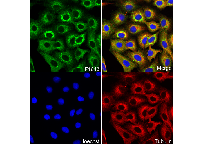 Immunofluorescent analysis of A549 cells using F1643 (green, 1:100), Hoechst (blue) and tubulin (Red).