|
Hoe te citeren 1. Voor in-tekst citatie (materialen en methoden): 2. Voor de tabel met belangrijke bronnen: |
||
|
Gratis nummer: (877) 796-6397 -- Alleen VS en Canada -- |
Fax: +1-832-582-8590 Bestellingen: +1-832-582-8158 |
Technische ondersteuning: +1-832-582-8158 Ext:3 Vermeld uw bestelnummer in de e-mail. Wij streven ernaar alle e-mailvragen binnen één werkdag te beantwoorden. |
Biologische Beschrijving
| Specificiteit | Collagen IV Antibody [E5G18] detecteert endogene niveaus van totaal Collagen IV-eiwit. |
|---|---|
| Achtergrond | Collagen IV is het primaire structurele collageen van basale membranen, gespecialiseerde extracellulaire matrices die mechanische ondersteuning bieden, celadhesie reguleren en andere matrixcomponenten organiseren. Het is een heterotrimeer dat bestaat uit combinaties van zes α-ketens (α1–α6) gecodeerd door verschillende genen, typisch gerangschikt als twee α1- en één α2-keten in de meeste weefsels, intracellulair geassembleerd tot triple-helix protomeren. Deze protomeren worden afgescheiden en gecrosslinkt via hun NC1- en 7S-domeinen om een stabiel, plaatachtig netwerk te vormen dat lamininen, proteoglycanen en groeifactoren verankert. Collagen IV wordt breed tot expressie gebracht in epitheliale en endotheliale basale membranen, met weefselspecifieke isoformensamenstelling, en speelt essentiële rollen in ontwikkeling, weefselintegriteit, celmigratie en Signaling via Integrin zoals α1β1 en α2β1. Defecten of afwijkende crosslinking van Collagen IV dragen bij aan diverse pathologieën, waaronder het Alport-syndroom, de ziekte van Goodpasture, diabetische nefropathie en bepaalde dermatologische aandoeningen. |
Gebruiksinformatie
| Toepassing | IHC | Verdunning |
|
||
|---|---|---|---|---|---|
| Reactiviteit | Human | ||||
| Bron | Mouse Monoclonal Antibody | MW | |||
| Opslagbuffer | PBS, pH 7.2+50% Glycerol+0.05% BSA+0.01% NaN3 | Opslag (Vanaf de datum van ontvangst) |
-20°C (avoid freeze-thaw cycles), 2 years | ||
| IHC |
Experimental Protocol:
Deparaffinization/Rehydration
1. Deparaffinize/hydrate sections:
2. Incubate sections in three washes of xylene for 5 min each.
3. Incubate sections in two washes of 100% ethanol for 10 min each.
4. Incubate sections in two washes of 95% ethanol for 10 min each.
5. Wash sections two times in dH2O for 5 min each.
6.Antigen retrieval: For Citrate: Heat slides in a microwave submersed in 1X citrate unmasking solution until boiling is initiated; continue with 10 min at a sub-boiling temperature (95°-98°C). Cool slides on bench top for 30 min.
Staining
1. Wash sections in dH2O three times for 5 min each.
2. Incubate sections in 3% hydrogen peroxide for 10 min.
3. Wash sections in dH2O two times for 5 min each.
4. Wash sections in wash buffer for 5 min.
5. Block each section with 100–400 µl of blocking solution for 1 hr at room temperature.
6. Remove blocking solution and add 100–400 µl primary antibody diluent in to each section. Incubate overnight at 4°C.
7. Remove antibody solution and wash sections with wash buffer three times for 5 min each.
8. Cover section with 1–3 drops HRPas needed. Incubate in a humidified chamber for 30 min at room temperature.
9. Wash sections three times with wash buffer for 5 min each.
10. Add DAB Chromogen Concentrate to DAB Diluent and mix well before use.
11. Apply 100–400 µl DAB to each section and monitor closely. 1–10 min generally provides an acceptable staining intensity.
12. Immerse slides in dH2O.
13. If desired, counterstain sections with hematoxylin.
14. Wash sections in dH2O two times for 5 min each.
15. Dehydrate sections: Incubate sections in 95% ethanol two times for 10 sec each; Repeat in 100% ethanol, incubating sections two times for 10 sec each; Repeat in xylene, incubating sections two times for 10 sec each.
16. Mount sections with coverslips and mounting medium.
|
Referenties
|
Toepassingsgegevens
IHC
Gevalideerd door Selleck
-
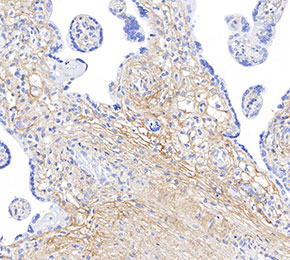 Immunohistochemical analysis of formalin fixed paraffin embedded human placenta tissue with F1667 at 1:125 dilution.