|
Hoe te citeren 1. Voor in-tekst citatie (materialen en methoden): 2. Voor de tabel met belangrijke bronnen: |
||
|
Gratis nummer: (877) 796-6397 -- Alleen VS en Canada -- |
Fax: +1-832-582-8590 Bestellingen: +1-832-582-8158 |
Technische ondersteuning: +1-832-582-8158 Ext:3 Vermeld uw bestelnummer in de e-mail. Wij streven ernaar alle e-mailvragen binnen één werkdag te beantwoorden. |
Biologische Beschrijving
| Specificiteit | ATG4B Antibody [P14M3] detecteert endogene niveaus van totaal ATG4B-eiwit. |
|---|---|
| Achtergrond | ATG4B is een cruciale cysteïne-protease bij Autophagy, verantwoordelijk voor de verwerking en recycling van ATG8-familie-eiwitten zoals LC3 en GABARAP, waardoor de biogenese van autofagosomen bij eukaryoten mogelijk wordt. ATG4B bevat een katalytische triade bestaande uit Cys74, Asp278 en His280 binnen een papaïne-achtige vouwing, met een regulerende lus en Trp142 die het enzym in een autogeïnhibeerde toestand houden totdat substraatbinding conformationele veranderingen induceert voor peptidebindinghydrolyse. N-terminale motieven vergroten verder de specificiteit voor LC3 geconjugeerd aan fosfatidylethanolamine (PE) op autofagische membranen. De primaire functie van ATG4B is het blootleggen van de C-terminale glycine op pro-LC3 door een initiële primingsplitsing, waardoor het wordt voorbereid op conjugatie aan PE via de ATG7/ATG3 ubiquitine-achtige cascade, en vervolgens het katalyseren van de delipidatie van LC3-PE op het buitenste autofagosomale membraan, waardoor de recycling van vrij LC3 wordt vergemakkelijkt. Met de breedste substraatselectiviteit onder de ATG4-paralogen, geeft ATG4B voornamelijk de voorkeur aan LC3, waardoor de elongatie van het autofagosoommembraan, de verankering en de autofagische flux in evenwicht worden gehouden. De activiteit wordt strak gereguleerd door redoxmodificaties aan Cys292 en Cys361 en door fosforylering, waardoor Autophagy wordt aangepast aan cellulaire stress; overexpressie van wildtype ATG4B kan LC3-lipidatie blokkeren door overmatige deconjugatie, terwijl katalytisch inactieve mutanten zoals C74A LC3 sequestreren en Autophagy remmen. Deregulatie van ATG4B-activiteit is gekoppeld aan tumorigenese als gevolg van een verminderde autofagische flux. |
Gebruiksinformatie
| Toepassing | WB, FCM | Verdunning |
|
||||
|---|---|---|---|---|---|---|---|
| Reactiviteit | Mouse, Human | ||||||
| Bron | Rabbit Monoclonal Antibody | MW | 44 kDa | ||||
| Opslagbuffer | PBS, pH 7.2+50% Glycerol+0.05% BSA+0.01% NaN3 | Opslag (Vanaf de datum van ontvangst) |
-20°C (avoid freeze-thaw cycles), 2 years | ||||
| WB |
Experimental Protocol:
Sample preparation
1. Tissue: Lyse the tissue sample by adding an appropriate volume of ice-cold RIPA/NP-40 Lysis Buffer (containing Protease Inhibitor Cocktail),and homogenize the tissue at a low temperature. 2. Adherent cell: Aspirate the culture medium and wash the cells with ice-cold PBS twice. Lyse the cells by adding an appropriate volume of RIPA/NP-40 Lysis Buffer (containing Protease Inhibitor Cocktail) and put the sample on ice for 5 min. 3. Suspension cell: Transfer the culture medium to a pre-cooled centrifuge tube. Centrifuge and aspirate the supernatant. Wash the cells with ice-cold PBS twice. Lyse the cells by adding an appropriate volume of RIPA/NP-40 Lysis Buffer (containing Protease Inhibitor Cocktail) and put the sample on ice for 5 min. 4. Place the lysate into a pre-cooled microcentrifuge tube. Centrifuge at 4°C for 15 min. Collect the supernatant;
5. Remove a small volume of lysate to determine the protein concentration;
6. Combine the lysate with protein loading buffer. Boil 20 µL sample under 95-100°C for 5 min. Centrifuge for 5 min after cool down on ice.
Electrophoretic separation
1. According to the concentration of extracted protein, load appropriate amount of protein sample and marker onto SDS-PAGE gels for electrophoresis. Recommended separating gel (lower gel) concentration: 10%. Reference Table for Selecting SDS-PAGE Separation Gel Concentrations 2. Power up 80V for 30 minutes. Then the power supply is adjusted (110 V~150 V), the Marker is observed, and the electrophoresis can be stopped when the indicator band of the predyed protein Marker where the protein is located is properly separated. (Note that the current should not be too large when electrophoresis, too large current (more than 150 mA) will cause the temperature to rise, affecting the result of running glue. If high currents cannot be avoided, an ice bath can be used to cool the bath.)
Transfer membrane
1. Take out the converter, soak the clip and consumables in the pre-cooled converter;
2. Activate PVDF membrane with methanol for 1 min and rinse with transfer buffer;
3. Install it in the order of "black edge of clip - sponge - filter paper - filter paper - glue -PVDF membrane - filter paper - filter paper - sponge - white edge of clip"; 4. The protein was electrotransferred to PVDF membrane. ( 0.45 µm PVDF membrane is recommended ) Reference Table for Selecting PVDF Membrane Pore Size Specifications Recommended conditions for wet transfer: 200 mA, 120 min. ( Note that the transfer conditions can be adjusted according to the protein size. For high-molecular-weight proteins, a higher current and longer transfer time are recommended. However, ensure that the transfer tank remains at a low temperature to prevent gel melting.)
Block
1. After electrotransfer, wash the film with TBST at room temperature for 5 minutes;
2. Incubate the film in the blocking solution for 1 hour at room temperature;
3. Wash the film with TBST for 3 times, 5 minutes each time.
Antibody incubation
1. Use 5% skim milk powder to prepare the primary antibody working liquid (recommended dilution ratio for primary antibody 1:1000), gently shake and incubate with the film at 4°C overnight; 2. Wash the film with TBST 3 times, 5 minutes each time;
3. Add the secondary antibody to the blocking solution and incubate with the film gently at room temperature for 1 hour;
4. After incubation, wash the film with TBST 3 times for 5 minutes each time.
Antibody staining
1. Add the prepared ECL luminescent substrate (or select other color developing substrate according to the second antibody) and mix evenly;
2. Incubate with the film for 1 minute, remove excess substrate (keep the film moist), wrap with plastic film, and expose in the imaging system.
|
Referenties
|
Toepassingsgegevens
WB
Gevalideerd door Selleck
-
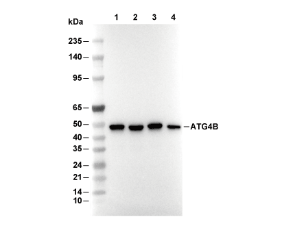 Lane 1: Ramos, Lane 2: 293T, Lane 3: Jurkat, Lane 4: Hela