|
Hoe te citeren 1. Voor in-tekst citatie (materialen en methoden): 2. Voor de tabel met belangrijke bronnen: |
||
|
Gratis nummer: (877) 796-6397 -- Alleen VS en Canada -- |
Fax: +1-832-582-8590 Bestellingen: +1-832-582-8158 |
Technische ondersteuning: +1-832-582-8158 Ext:3 Vermeld uw bestelnummer in de e-mail. Wij streven ernaar alle e-mailvragen binnen één werkdag te beantwoorden. |
Biologische Beschrijving
| Specificiteit | Artemis Antibody [H20J21] detecteert endogene niveaus van totaal Artemis-eiwit. |
|---|---|
| Achtergrond | Artemis (DCLRE1C, ook bekend als SNM1C) is een ubiquitair tot expressie gebrachte nucleaire nuclease die behoort tot de metallo-β-lactamase (MBL) superfamilie, essentieel voor niet-homologe eindverbinding (NHEJ) DNA-reparatie en V(D)J-recombinatie in ontwikkelende lymfocyten. Artemis is een eiwit van 692 aminozuren met een N-terminaal katalytisch domein (residuen 1-360) bestaande uit een kern MBL-vouw (α/β-β/α sandwich) en een ingebed β-CASP-subdomein dat geconserveerde motieven 1-4 (His33, His35, His115, Asp116 die Zn²⁺-ionen M1/M2 coördineren) en motieven A-C voor nucleïnezuurverwerking bevat, plus een grotendeels ongestructureerde C-terminale regulerende staart (residuen 361-692) die de activiteit auto-inhibeert en de DNA-PKcs-verankering medieert. DNA-PKcs fosforyleert Artemis (bijv. bij Thr127, Ser251) na DSB-herkenning, waardoor de structuurspecifieke endonuclease-activiteit wordt geactiveerd om haarspeld-verzegelde coderende uiteinden te openen in V(D)J-recombinatie, 5'/3' overhangs, flaps en bubbels in NHEJ te trimmen voor XRCC4/LIG4-ligatie, en IR-geïnduceerde complexe uiteinden te verwerken terwijl HR wordt tegengegaan via 53BP1-PTIP-interactie. ATM/ATR fosforyleren Artemis verder voor G2/M- en S-fase checkpointherstel, waardoor Cdk1-cycline B-activatie en cycline E-degradatie via SCF^{Fbw7} worden bevorderd. Biallelische mutaties veroorzaken radiosensitieve ernstige gecombineerde immuundeficiëntie (RS-SCID) met defecte V(D)J-verbinding en genoominstabiliteit die predisponeert voor maligniteit. |
Gebruiksinformatie
| Toepassing | WB, IP | Verdunning |
|
||||
|---|---|---|---|---|---|---|---|
| Reactiviteit | Human, Monkey | ||||||
| Bron | Rabbit Monoclonal Antibody | MW | 90 kDa | ||||
| Opslagbuffer | PBS, pH 7.2+50% Glycerol+0.05% BSA+0.01% NaN3 | Opslag (Vanaf de datum van ontvangst) |
-20°C (avoid freeze-thaw cycles), 2 years | ||||
| WB |
Experimental Protocol:
Sample preparation
1. Tissue: Lyse the tissue sample by adding an appropriate volume of ice-cold RIPA/Nuclear Lysis Buffer (containing Protease Inhibitor Cocktail),and homogenize the tissue at a low temperature. 2. Adherent cell: Aspirate the culture medium and wash the cells with ice-cold PBS twice. Lyse the cells by adding an appropriate volume of RIPA/Nuclear Lysis Buffer (containing Protease Inhibitor Cocktail) and put the sample on ice for 5 min. 3. Suspension cell: Transfer the culture medium to a pre-cooled centrifuge tube. Centrifuge and aspirate the supernatant. Wash the cells with ice-cold PBS twice. Lyse the cells by adding an appropriate volume of RIPA/Nuclear Lysis Buffer (containing Protease Inhibitor Cocktail) and put the sample on ice for 5 min. 6. Add protein loading buffer, heat 20 μL of the sample at 95~100°C for 5 min, let it cool down on ice and then centrifuge for 5 min. Electrophoretic separation
1. According to the concentration of extracted protein, load appropriate amount of protein sample and marker onto SDS-PAGE gels for electrophoresis. Recommended separating gel (lower gel) concentration: 10%. Reference Table for Selecting SDS-PAGE Separation Gel Concentrations 2. Power up 80V for 30 minutes. Then the power supply is adjusted (110 V~150 V), the Marker is observed, and the electrophoresis can be stopped when the indicator band of the predyed protein Marker where the protein is located is properly separated. (Note that the current should not be too large when electrophoresis, too large current (more than 150 mA) will cause the temperature to rise, affecting the result of running glue. If high currents cannot be avoided, an ice bath can be used to cool the bath.)
Transfer membrane
1. Take out the converter, soak the clip and consumables in the pre-cooled converter;
2. Activate PVDF membrane with methanol for 1 min and rinse with transfer buffer;
3. Install it in the order of "black edge of clip - sponge - filter paper - filter paper - glue -PVDF membrane - filter paper - filter paper - sponge - white edge of clip"; 4. The protein was electrotransferred to PVDF membrane. ( 0.45 µm PVDF membrane is recommended ) Reference Table for Selecting PVDF Membrane Pore Size Specifications Recommended conditions for wet transfer: 200 mA, 120 min. ( Note that the transfer conditions can be adjusted according to the protein size. For high-molecular-weight proteins, a higher current and longer transfer time are recommended. However, ensure that the transfer tank remains at a low temperature to prevent gel melting.)
Block
1. After electrotransfer, wash the film with TBST at room temperature for 5 minutes;
2. Incubate the film in the blocking solution for 1 hour at room temperature;
3. Wash the film with TBST for 3 times, 5 minutes each time.
Antibody incubation
1. Use 5% skim milk powder to prepare the primary antibody working liquid (recommended dilution ratio for primary antibody 1:1000), gently shake and incubate with the film at 4°C overnight; 2. Wash the film with TBST 3 times, 5 minutes each time;
3. Add the secondary antibody to the blocking solution and incubate with the film gently at room temperature for 1 hour;
4. After incubation, wash the film with TBST 3 times for 5 minutes each time.
Antibody staining
1. Add the prepared ECL luminescent substrate (or select other color developing substrate according to the second antibody) and mix evenly;
2. Incubate with the film for 1 minute, remove excess substrate (keep the film moist), wrap with plastic film, and expose in the imaging system. (Exposure time of at least 60s is recommended)
|
Referenties
|
Toepassingsgegevens
WB
Gevalideerd door Selleck
-
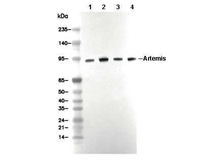 Lane 1: Jurkat, Lane 2: HT-29, Lane 3: PC-3, Lane 4: MCF-7