|
Hoe te citeren 1. Voor in-tekst citatie (materialen en methoden): 2. Voor de tabel met belangrijke bronnen: |
||
|
Gratis nummer: (877) 796-6397 -- Alleen VS en Canada -- |
Fax: +1-832-582-8590 Bestellingen: +1-832-582-8158 |
Technische ondersteuning: +1-832-582-8158 Ext:3 Vermeld uw bestelnummer in de e-mail. Wij streven ernaar alle e-mailvragen binnen één werkdag te beantwoorden. |
Biologische Beschrijving
| Specificiteit | Aldolase + Aldolase B + Aldolase C Antibody [F19A18] herkent endogene niveaus van totaal Aldolase + Aldolase B + Aldolase C-eiwit. |
|---|---|
| Achtergrond | Aldolase + Aldolase B + Aldolase C zijn belangrijke glycolytische enzymen die de reversibele splitsing van fructose-1,6-bisfosfaat of fructose-1-fosfaat in dihydroxyacetonfosfaat en glyceraldehyde-3-fosfaat of glyceraldehyde katalyseren. Bij gewervelde dieren delen drie aldolase-iso-enzymen – aldolase A, B en C – een sterk geconserveerde αβ-barrelvouw en een katalytisch mechanisme, maar verschillen ze in weefseldistributie en functionele rollen. Aldolase A wordt voornamelijk tot expressie gebracht in spieren en rode bloedcellen, ter ondersteuning van een hoge glycolytische flux; aldolase B wordt voornamelijk gevonden in de lever, nieren en dunne darm, waar het cruciale rollen speelt in zowel glycolyse als gluconeogenese; en aldolase C is verrijkt in de hersenen en neurale weefsels, waar de exacte metabole functie minder gedefinieerd blijft, maar mogelijk verband houdt met neuro-ontwikkeling en energiemetabolisme. Ondanks hun vergelijkbare actieve-site-architectuur en gedeelde katalytische residuen, verschillen de iso-enzymen in iso-enzymspecifieke residuen (ISRs), die clusteren op het eiwitoppervlak en mogelijk verschillende eiwit-eiwitinteracties of regulerende eigenschappen mediëren. Alle drie aldolasen vertonen ook niet-katalytische "moonlighting"-functies, waarbij ze op een weefselspecifieke manier interageren met cytoskeletale eiwitten, metabole enzymen en andere macromoleculen, waardoor hun cellulaire rollen verder worden gediversifieerd. |
Gebruiksinformatie
| Toepassing | WB, FCM | Verdunning |
|
||||
|---|---|---|---|---|---|---|---|
| Reactiviteit | Human, Mouse, Rat, Pig | ||||||
| Bron | Rabbit Monoclonal Antibody | MW | 39 kDa | ||||
| Opslagbuffer | PBS, pH 7.2+50% Glycerol+0.05% BSA+0.01% NaN3 | Opslag (Vanaf de datum van ontvangst) |
-20°C (avoid freeze-thaw cycles), 2 years | ||||
| WB |
Experimental Protocol:
Sample preparation
1. Tissue: Lyse the tissue sample by adding an appropriate volume of ice-cold RIPA/NP-40 Lysis Buffer (containing Protease Inhibitor Cocktail),and homogenize the tissue at a low temperature. 2. Adherent cell: Aspirate the culture medium and wash the cells with ice-cold PBS twice. Lyse the cells by adding an appropriate volume of RIPA/NP-40 Lysis Buffer (containing Protease Inhibitor Cocktail) and put the sample on ice for 5 min. 3. Suspension cell: Transfer the culture medium to a pre-cooled centrifuge tube. Centrifuge and aspirate the supernatant. Wash the cells with ice-cold PBS twice. Lyse the cells by adding an appropriate volume of RIPA/NP-40 Lysis Buffer (containing Protease Inhibitor Cocktail) and put the sample on ice for 5 min. 4. Place the lysate into a pre-cooled microcentrifuge tube. Centrifuge at 4°C for 15 min. Collect the supernatant;
5. Remove a small volume of lysate to determine the protein concentration;
6. Combine the lysate with protein loading buffer. Boil 20 µL sample under 95-100°C for 5 min. Centrifuge for 5 min after cool down on ice.
Electrophoretic separation
1. According to the concentration of extracted protein, load appropriate amount of protein sample and marker onto SDS-PAGE gels for electrophoresis. Recommended separating gel (lower gel) concentration: 10%. Reference Table for Selecting SDS-PAGE Separation Gel Concentrations 2. Power up 80V for 30 minutes. Then the power supply is adjusted (110 V~150 V), the Marker is observed, and the electrophoresis can be stopped when the indicator band of the predyed protein Marker where the protein is located is properly separated. (Note that the current should not be too large when electrophoresis, too large current (more than 150 mA) will cause the temperature to rise, affecting the result of running glue. If high currents cannot be avoided, an ice bath can be used to cool the bath.)
Transfer membrane
1. Take out the converter, soak the clip and consumables in the pre-cooled converter;
2. Activate PVDF membrane with methanol for 1 min and rinse with transfer buffer;
3. Install it in the order of "black edge of clip - sponge - filter paper - filter paper - glue -PVDF membrane - filter paper - filter paper - sponge - white edge of clip"; 4. The protein was electrotransferred to PVDF membrane. ( 0.45 µm PVDF membrane is recommended ) Reference Table for Selecting PVDF Membrane Pore Size Specifications Recommended conditions for wet transfer: 200 mA, 60 min. ( Note that the transfer conditions can be adjusted according to the protein size. For high-molecular-weight proteins, a higher current and longer transfer time are recommended. However, ensure that the transfer tank remains at a low temperature to prevent gel melting.)
Block
1. After electrotransfer, wash the film with TBST at room temperature for 5 minutes;
2. Incubate the film in the blocking solution for 1 hour at room temperature;
3. Wash the film with TBST for 3 times, 5 minutes each time.
Antibody incubation
1. Use 5% skim milk powder to prepare the primary antibody working liquid (recommended dilution ratio for primary antibody 1:1000), gently shake and incubate with the film at 4°C overnight; 2. Wash the film with TBST 3 times, 5 minutes each time;
3. Add the secondary antibody to the blocking solution and incubate with the film gently at room temperature for 1 hour;
4. After incubation, wash the film with TBST 3 times for 5 minutes each time.
Antibody staining
1. Add the prepared ECL luminescent substrate (or select other color developing substrate according to the second antibody) and mix evenly;
2. Incubate with the film for 1 minute, remove excess substrate (keep the film moist), wrap with plastic film, and expose in the imaging system.
|
Referenties
|
Toepassingsgegevens
WB
Gevalideerd door Selleck
-
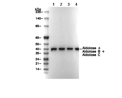 Lane 1: Hela, Lane 2: Mouse brain, Lane 3: Rat brain, Lane 4: Pig heart