Technische gegevens
| Formule | C15H14N2O4S |
||||||
| Moleculair gewicht | 318.35 | CAS-nr. | 866323-14-0 | ||||
| Oplosbaarheid (25°C)* | In vitro | DMSO | 64 mg/mL (201.03 mM) | ||||
| Water | Insoluble | ||||||
| Ethanol | Insoluble | ||||||
| In vivo (Voeg oplosmiddelen afzonderlijk en in volgorde toe aan het product.) |
|
||||||
|
* <1 mg/ml betekent licht oplosbaar of onoplosbaar. * Houd er rekening mee dat Selleck de oplosbaarheid van alle verbindingen intern test en de werkelijke oplosbaarheid enigszins kan afwijken van gepubliceerde waarden. Dit is normaal en is te wijten aan lichte batch-tot-batch variaties. * Verzending op kamertemperatuur (Stabiliteitstests tonen aan dat dit product zonder koelmaatregelen kan worden verzonden.) |
|||||||
Voorbereiden van stamoplossingen
Biologische activiteit
| Beschrijving | Belinostat is een nieuwe HDAC-remmer met een IC50 van 27 nM in een celvrije assay, met activiteit aangetoond in cisplatin-resistente tumoren. Belinostat (PXD101) induceert autophagy. | ||
|---|---|---|---|
| Doelen |
|
||
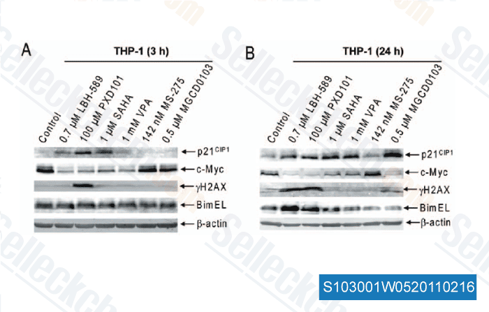
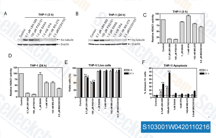
| In vitro | Belinostat remt de groei van tumorcellen (A2780, HCT116, HT29, WIL, CALU-3, MCF7, PC3 en HS852) met IC50 van 0,2-0,66 μM. PD101 vertoont lage activiteit in A2780/cp70 en 2780AD cellen, dit zijn cisplatin- en doxorubicine-resistente derivaten van A2780 cellen. Deze verbinding kan apoptose induceren via PARP-splitsing en acetylering van histonen H3/H4. Het remt de groei van blaaskankercellen, vooral in 5637 cellen, die een accumulatie van de G0-G1 fase, een afname van de S-fase en een toename van de G2-M fase vertonen. De groeiremmende activiteit van deze chemische stof op cellijnen wordt niet sterk beïnvloed door het multidrug-resistente fenotype, terwijl de activiteit van docetaxel duidelijk wordt beïnvloed. Het kan de groeiremmende activiteit van docetaxel of carboplatine in OVCAR-3 en A2780 cellen versterken. Deze verbinding vertoont ook verhoogde tubuline-acetylering in eierstokkankercellijnen. Een recente studie toont aan dat het proteïnekinase A activeert via een TGF-β-afhankelijk mechanisme en het survivin-mRNA verlaagt. | ||
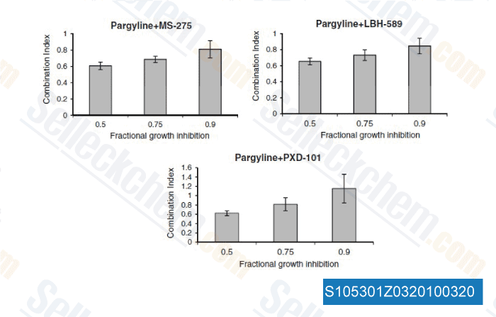
| In vivo | Belinostat veroorzaakt een significante vertraging van de tumorgroei in A2780 en A2780/cp70 xenograften bij een dosis van 10 mg/kg zonder effecten op het lichaamsgewicht. Deze verbinding induceert ook p21WAF1, HDAC core en celcommunicatiegenen in muizenblaastumoren. De monotherapie induceert dosis-proportionele antitumorale effecten met een TGI van 47% bij een dosis van 100 mg/kg in A2780 xenograft. De combinatie van deze chemische stof (100 mg/kg) met carboplatine (40 mg/kg) kan de tumorgroei vertragen van 18,6 dagen naar 22,5 dagen. In combinatie met bortezomib resulteert het in een grote tumorremming en gastro-intestinale toxiciteit bij muizen met bortezomib-resistente UMSCC-11A xenograft. | ||
| Kenmerken | Hoofdverbinding van Topotarget. |
Protocol (uit referentie)
| Kinase-assay:[1] |
|
|---|---|
| Celassay:[1] |
|
| Dierstudie:[1] |
|
Referenties
|
Klantproductvalidatie

-
Gegevens van [ Breast Cancer Res Treat , 2012 , 131, 777-789 ]
-
Gegevens van [ PLoS One , 2011 , 6, e17138 ]
-
Gegevens van [ PLoS One , 2011 , 6, e17138 ]
-
Gegevens van [ Breast Cancer Res Treat , 2010 , 131(3), 777-789 ]
Sellecks Belinostat (PXD101) Is geciteerd door 108 Publicaties
| A patient-derived T cell lymphoma biorepository uncovers pathogenetic mechanisms and host-related therapeutic vulnerabilities [ Cell Rep Med, 2025, S2666-3791(25)00102-8] | PubMed: 40147445 |
| Deacetylation of TALDO1 by HDAC6 promotes glycolysis and nasopharyngeal carcinoma progression through a moonlighting function [ Cell Death Dis, 2025, 16(1):743] | PubMed: 41120289 |
| The anticancer effect of the HDAC inhibitor belinostat is enhanced by inhibitors of Bcl-xL or Mcl-1 in ovarian cancer [ Mol Oncol, 2025, 10.1002/1878-0261.70050] | PubMed: 40483575 |
| Targeting the akt/mtor signaling pathway by maprotiline leads to tumor suppression in T-cell lymphoma [ Ann Hematol, 2025, 10.1007/s00277-025-06571-z] | PubMed: 40892074 |
| Role of the NuRD complex and altered proteostasis in cancer cell quiescence [ bioRxiv, 2025, 2025.02.10.637435] | PubMed: 39990343 |
| Orthogonal proteogenomic analysis identifies the druggable PA2G4-MYC axis in 3q26 AML [ Nat Commun, 2024, 15(1):4739] | PubMed: 38834613 |
| Interferon-induced factor 16 is essential in metastatic melanoma to maintain STING levels and the immune responses upon IFN-γ response pathway activation [ J Immunother Cancer, 2024, 12(10)e009590] | PubMed: 39424359 |
| Chronic hypoxia stabilizes 3βHSD1 via autophagy suppression [ Cell Rep, 2024, 43(1):113575] | PubMed: 38181788 |
| PXD101 inhibits malignant progression and radioresistance of glioblastoma by upregulating GADD45A [ J Transl Med, 2024, 22(1):1047] | PubMed: 39568000 |
| Establishment, characterization, and biobanking of 36 pancreatic cancer organoids: prediction of metastasis in resectable pancreatic cancer [ Cell Oncol (Dordr), 2024, 10.1007/s13402-024-00939-5] | PubMed: 38619751 |
RETOURBELEID
Selleck Chemicals onvoorwaardelijke retourbeleid zorgt voor een soepele online winkelervaring voor onze klanten. Als u op enigerlei wijze ontevreden bent met uw aankoop, kunt u elk artikel(en) binnen 7 dagen na ontvangst retourneren. In geval van problemen met de productkwaliteit, zowel protocolgerelateerde als productgerelateerde problemen, kunt u elk artikel(en) binnen 365 dagen na de oorspronkelijke aankoopdatum retourneren. Volg de onderstaande instructies bij het retourneren van producten.
VERZENDING EN OPSLAG
Selleck producten worden bij kamertemperatuur vervoerd. Als u het product op kamertemperatuur ontvangt, wees dan gerust, de Selleck kwaliteitsinspectieafdeling heeft experimenten uitgevoerd om te controleren of de normale temperatuurplaatsing van één maand de biologische activiteit van poederproducten niet beïnvloedt. Na ontvangst dient u het product op te slaan volgens de vereisten beschreven in het gegevensblad. De meeste Selleck producten zijn stabiel onder de aanbevolen omstandigheden.
NIET VOOR HUMANE, VETERINAIRE DIAGNOSTISCHE OF THERAPEUTISCHE DOELEINDEN.